


Case #186 - August, 2006
 ShareCompartir
A man went to a local hospital with abdominal pain and weight loss. He reported that he frequently travels to South America and had previously been diagnosed with ascariasis, although recent stool specimens were negative. The laboratory saw the objects in Figures A, B, and C in the patient’s stool samples. The objects measured 10 to 15 µm. Figure A was taken at 400× magnification from wet mount stained with iodine. Figures B and C were taken at 1000× magnification from a trichrome stained slide. What is your diagnosis? Based on what criteria?
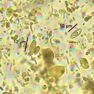
Figure A

Figure B
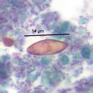
Figure C
Case Answer
The objects are artifacts, probably plant material, and a diagnosis was given of No Parasites Found (NPF). Although the objects were morphologically similar to Cystoisospora oocysts (Figure D), they were smaller than Cystoisospora, which ranges from 25 to 30 µm.

Figure A
More on: Artifacts
This case was kindly contributed by the Wisconsin State Laboratory of Hygiene.
Images presented in the monthly case studies are from specimens submitted for diagnosis or archiving. On rare occasions, clinical histories given may be partly fictitious.
DPDx is an education resource designed for health professionals and laboratory scientists. For an overview including prevention and control visit www.cdc.gov/parasites/.
- Page last reviewed: August 24, 2016
- Page last updated: August 24, 2016
- Content source:
- Global Health – Division of Parasitic Diseases and Malaria
- Notice: Linking to a non-federal site does not constitute an endorsement by HHS, CDC or any of its employees of the sponsors or the information and products presented on the site.
- Maintained By: