


Case #183 - July, 2006
 ShareCompartir
A family visited different U.S. states during a one week vacation. Approximately eight days after their trip, two family members began experiencing diarrhea. They went to their family physician and reported that while they were traveling they swam in hotel pools and at a waterpark. The physician requested stool samples for an ova and parasite (O & P) examination, along with other testing. The samples collected for the O & P were preserved in 10% formalin and then sent for examination. Figure A shows rounded objects seen on a modified acid-fast stained slide made from a formalin-ethyl acetate (FEA) concentrate of one of the specimens. The same objects were seen in specimens from the other family member who was ill. The objects measured 4.5-5.5 micrometers in diameter. What is your diagnosis? Based on what criteria?
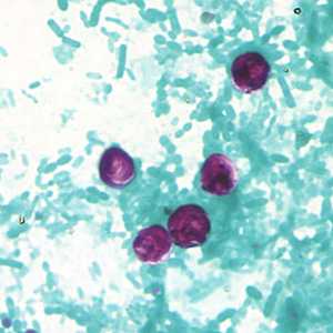
Figure A
Case Answer
This was a case of cryptosporidiosis caused by Cryptosporidium sp. Although many laboratories may only perform wet mounts and trichrome staining for an O & P, confirmatory identification of Cryptosporidium must include examination of acid-fast smears and/or DFA. In wet mounts, Cryptosporidium sp. oocysts can easily be confused with yeast cells and often numbers can be quite low making detection a challenge. Diagnostic features observed in the acid-fast stained smears were:
- the size of the oocysts (4-5 micrometers in diameter), which was consistent with oocysts of Cryptosporidium spp. that can infect humans.
- oocysts that stain positive with acid-fast stain, although Cryptosporidium oocysts may stain variably.
- the presence of sporozoites (Figure A, arrow) within one of the oocysts. This characteristic is often easier to see on an acid-fast stained slide. Also in stained preparations dark granules of residual material may be present within some of the oocysts.
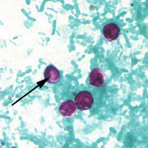
Figure A
Molecular tools are available to identify Cryptosporidium sp. at species level. Some of the species that infect humans cannot be distinguished morphologically.
More on: Cryptosporidiosis
Images presented in the monthly case studies are from specimens submitted for diagnosis or archiving. On rare occasions, clinical histories given may be partly fictitious.
DPDx is an education resource designed for health professionals and laboratory scientists. For an overview including prevention and control visit www.cdc.gov/parasites/.
- Page last reviewed: August 24, 2016
- Page last updated: August 24, 2016
- Content source:
- Global Health – Division of Parasitic Diseases and Malaria
- Notice: Linking to a non-federal site does not constitute an endorsement by HHS, CDC or any of its employees of the sponsors or the information and products presented on the site.
- Maintained By: