


Case #182 - June, 2006
 ShareCompartir
A researcher was studying parasites of public health concern found in snails and slugs. A small portion of one specimen had tissue removed and placed in a small dish with a HCl/pepsin solution. Many larvae were observed in the dish, however most were dead or dying, but a few larvae were active. Figure A shows one of the active larvae in at wet mount captured at 200× magnification. Figure B shows a larva under differential interference contrast (DIC) microscopy at 400× magnification. What is your identification of the objects? Based on what criteria?
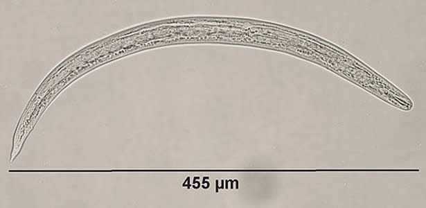
Figure A

Figure B
Case Answer
The nematode larvae were filariform third stage (L3) larvae of Angiostrongylus sp., the stage that is infective to mammals. Diagnostic features were:
- the length of the larvae, which were within range for Angiostrongylus sp. (425-524 micrometers). Hookworm filariform larvae are much longer (>800 micrometers), and one would not expect to find hookworm larvae contained in a snail or slug.
- the presence of chitinous rods called sclerotized rhabdions (see Figure A below, black arrows).
- a terminal projection on the tip of the tail which is characteristic of Angiostrongylus sp. (Figure B), although there is some overlap in size with filariform Strongyloides stercoralis, the latter’s tail is notched.
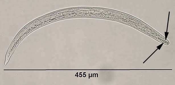
Figure A
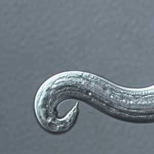
Figure B
Because it is difficult to differentiate L3 larvae of A. cantonensis from those of A. costaricensis based on morphology, epidemiologic data such as geographical location must also be relied on in order to make a diagnosis. Molecular analysis is useful to confirm the parasite at species level as well. In this specific case, DNA was extracted from a few larvae and a fragment of 1134 base pairs was amplified by PCR from the Angiostrongylus 18S rRNA gene. DNA sequencing analysis was performed on the amplified fragment, and the sequence showed 100% similarity with A. cantonensis 18S rRNA sequence deposited in GenBank under accession number AY295804.
More on: Angiostrongyliasis
Images presented in the monthly case studies are from specimens submitted for diagnosis or archiving. On rare occasions, clinical histories given may be partly fictitious.
DPDx is an education resource designed for health professionals and laboratory scientists. For an overview including prevention and control visit www.cdc.gov/parasites/.
- Page last reviewed: August 24, 2016
- Page last updated: August 24, 2016
- Content source:
- Global Health – Division of Parasitic Diseases and Malaria
- Notice: Linking to a non-federal site does not constitute an endorsement by HHS, CDC or any of its employees of the sponsors or the information and products presented on the site.
- Maintained By: