


Case #177 - April, 2006
 ShareCompartir
A 43-year-old man went to his physician complaining of fatigue. The man reported that he hiked and camped a lot throughout the northeastern United States weeks before the symptoms started. He also reported traveling to Kenya for vacation a month before his doctor’s visit and stated that he had complied with the recommended malaria prophylaxis regimen. A blood examination was ordered, and a thick and thin blood film were made and stained with Giemsa. Figure A shows what was seen on the thick blood film, and Figure B shows the thin blood film. What is your diagnosis? Based on what criteria?

Figure A
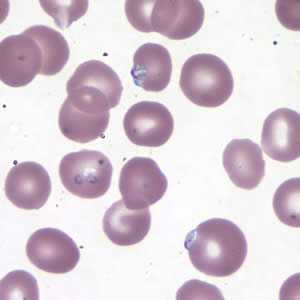
Figure B
Case Answer
This is a case of babesiosis caused by Babesia sp. Diagnostic features are:
- many ring-form organisms with red chromatin dots surrounded by blue cytoplasm. There is no yellow and/or brown pigment visible, which may be present in Plasmodium spp. Although the thick smear is useful in determining the presence of parasites, an examination of thin smears is necessary in order to make an accurate laboratory diagnosis.
- the presence of pleomorphic intra-erythrocytic parasites (Figure B), a feature consistent with Babesia spp.
- vacuolated ring-forms (Figure B below, black arrow).

Figure A
Additional testing methods, such as serology and/or molecular techniques, are always recommended to make a species identification of Babesia.
More on: Babesiosis
Images presented in the monthly case studies are from specimens submitted for diagnosis or archiving. On rare occasions, clinical histories given may be partly fictitious.
DPDx is an education resource designed for health professionals and laboratory scientists. For an overview including prevention and control visit www.cdc.gov/parasites/.
- Page last reviewed: August 24, 2016
- Page last updated: August 24, 2016
- Content source:
- Global Health – Division of Parasitic Diseases and Malaria
- Notice: Linking to a non-federal site does not constitute an endorsement by HHS, CDC or any of its employees of the sponsors or the information and products presented on the site.
- Maintained By: