


Case #176 - March, 2006
 ShareCompartir
A CDC microbiologist was looking through slide boxes that had been archived to determine whether the slide quality was good enough to continue storing them. He came across a pathology slide of heart tissue that was dated “1929.” Figures A-C show what was observed on the H & E stained preparation. What objects do you see in the images?

Figure A
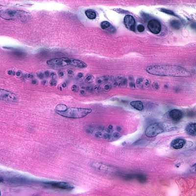
Figure B
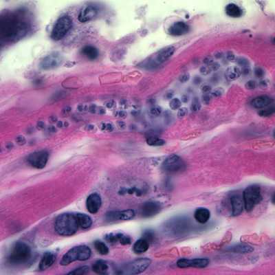
Figure C
Case Answer
The objects in the images are amastigotes. Determining the genus and species of a parasite is not possible based on morphologic features of amastigotes. The slide that was found was labeled, “Homo sapiens. Heart tissue infected with Trypanosoma cruzi, Chagas.” Diagnostic features included:
- the amastigote form of the parasite (Figures A-C, within black circles), identified by the presence of a nucleus and a distinct, sometimes rod-shaped, kinetoplast (yellow arrows).
- amastigotes that appear to be in the process of transforming into trypomastigotes (white circle, Figure A).

Figure A
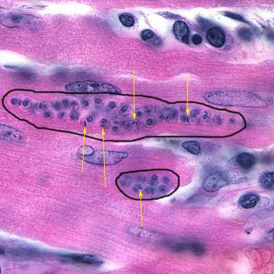
Figure B
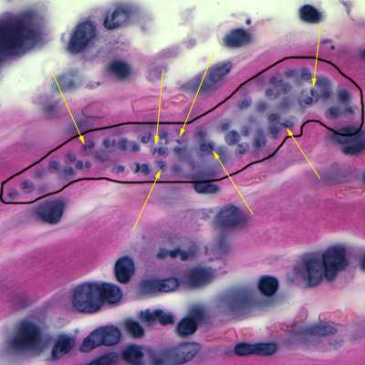
Figure C
More on: American trypanosomiasis
This case was kindly contributed by the Auckland City Hospital, New Zealand and images were taken by Dr. Allen Heath from AgResearch-Wallaceville Animal Research Centre.
Images presented in the monthly case studies are from specimens submitted for diagnosis or archiving. On rare occasions, clinical histories given may be partly fictitious.
DPDx is an education resource designed for health professionals and laboratory scientists. For an overview including prevention and control visit www.cdc.gov/parasites/.
- Page last reviewed: August 24, 2016
- Page last updated: August 24, 2016
- Content source:
- Global Health – Division of Parasitic Diseases and Malaria
- Notice: Linking to a non-federal site does not constitute an endorsement by HHS, CDC or any of its employees of the sponsors or the information and products presented on the site.
- Maintained By: