


Case #175 - March, 2006
 ShareCompartir
An elderly patient underwent a colonoscopy for polyps. A worm was seen near the appendiceal orifice and a biopsy was taken, fixed in formalin, and sent to histology. The sections were stained with hematoxylin and eosin (H & E). Figures A-C show what was observed on the slides at 100×, 400×, and 1000× magnification respectively. Objects such as the one shown in Figure C measured approximately 50 µm in length. What is your identification of this incidental finding? Based on what criteria?
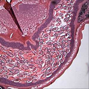
Figure A
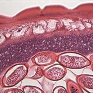
Figure B

Figure C
Case Answer
This was a case of trichuriasis, caused by Trichuris trichiura. Diagnostic morphologic features included:
- eggs in utero that were within the size range of T. trichiura (50 to 54 µm in length).
- the presence of polar plugs.
- barrel-shaped eggs, another typical characteristic of T. trichiura.
More on: Trichuriasis
This case was kindly contributed by the Oregon Public Health Laboratory.
Images presented in the monthly case studies are from specimens submitted for diagnosis or archiving. On rare occasions, clinical histories given may be partly fictitious.
DPDx is an education resource designed for health professionals and laboratory scientists. For an overview including prevention and control visit www.cdc.gov/parasites/.
- Page last reviewed: August 24, 2016
- Page last updated: August 24, 2016
- Content source:
- Global Health – Division of Parasitic Diseases and Malaria
- Notice: Linking to a non-federal site does not constitute an endorsement by HHS, CDC or any of its employees of the sponsors or the information and products presented on the site.
- Maintained By: