


Case #164 - September, 2005
 ShareCompartir
A 42-year-old animal trapper sought medical attention regarding an ulcerative lesion on his arm (Figure A). He reported that he had been in Bolivia two months ago and that the lesion on his arm had grown worse since his return. A biopsy of the lesion on his arm was performed and a touch prep smear prepared and stained with Giemsa. The tissue was sent to pathology to be sectioned and stained with H & E. Figures B and C show objects observed on the Giemsa stained touch prep smears, and Figures D and E show objects observed on one of the H & E slides. What is your diagnosis? Based on what criteria?
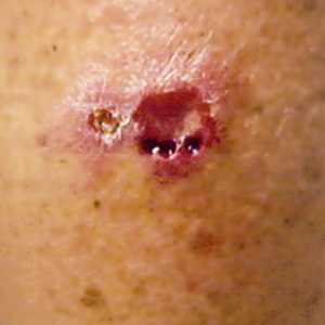
Figure A
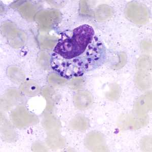
Figure B
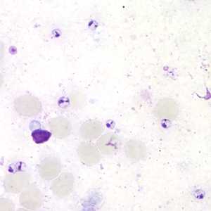
Figure C
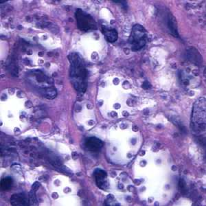
Figure D
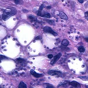
Figure E
Case Answer
This was a case of leishmaniasis caused by Leishmania sp. Genus and species determination should not be based on observation of amastigotes and/or lesion morphology alone. When travel history is compared to current knowledge of which species of Leishmania are found in Bolivia, this case was probably caused by L. braziliensis or L. amazonensis, both of which can progress to the mucocutaneous form. Techniques such as PCR or isoenzyme analysis can confirm the species of Leishmania causing disease. The patient in this case left the country before a follow-up biopsy could be done for further testing so a final species determination was not confirmed.
More on: Leishmaniasis
Images presented in the monthly case studies are from specimens submitted for diagnosis or archiving. On rare occasions, clinical histories given may be partly fictitious.
DPDx is an education resource designed for health professionals and laboratory scientists. For an overview including prevention and control visit www.cdc.gov/parasites/.
- Page last reviewed: August 24, 2016
- Page last updated: August 24, 2016
- Content source:
- Global Health – Division of Parasitic Diseases and Malaria
- Notice: Linking to a non-federal site does not constitute an endorsement by HHS, CDC or any of its employees of the sponsors or the information and products presented on the site.
- Maintained By: