


Case #156 - May, 2005
 ShareCompartir
A 25-year-old man living in South America became ill with intestinal cramping and watery diarrhea. Stool specimens were collected and an O & P exam was ordered by his physician. The lab performed an FEA concentration on the sample, prepared wet mounts, and examined them using UV fluorescence microscopy. Images were captured and submitted to DPDx/CDC. Figures A and B were taken at 400× magnification. What is your diagnosis? Based on what criteria?
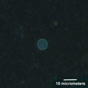
Figure A
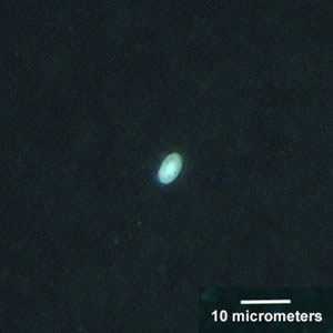
Figure B
Case Answer
This was a case of cyclosporiasis caused by Cyclospora sp. (most likely Cyclospora cayetanensis). The object in Figure B is an artifact. Diagnostic features were:
- the size of the object in Figure A and the autofluorescence of the oocyst wall, both of which were consistent with Cyclospora sp. Cyclospora spp. range in size from 8 to 10 micrometers.
There are numerous items that may fluoresce when using UV microscopy so careful observation of morphologic features under bright field microscopy is equally important when examining specimens (see February 2001’s case study 53 to see an image captured from bright field DIC microscopy). A secondary smear stained using modified Kinyoun’s acid-fast, or the hot safranin technique, is an alterative confirmatory technique, which also allows for a permanent record of the specimen.
More on: Cyclosporiasis
Images presented in the monthly case studies are from specimens submitted for diagnosis or archiving. On rare occasions, clinical histories given may be partly fictitious.
DPDx is an education resource designed for health professionals and laboratory scientists. For an overview including prevention and control visit www.cdc.gov/parasites/.
- Page last reviewed: August 24, 2016
- Page last updated: August 24, 2016
- Content source:
- Global Health – Division of Parasitic Diseases and Malaria
- Notice: Linking to a non-federal site does not constitute an endorsement by HHS, CDC or any of its employees of the sponsors or the information and products presented on the site.
- Maintained By: