


Case #149 - February, 2005
 ShareCompartir
A live worm was recovered from a 3-year-old’s diaper. The child was healthy and had not traveled outside of the country. The worm was preserved in 10% formaldehyde and forwarded to the Swedish Institute for Infectious Disease Control (SMI) for examination. The worm’s size was reportedly 6 cm long when first discovered, although when fixed, it measured 3.5 cm in length and 0.6 cm in width. Also due to fixation, the worm was brittle and had almost broke in half; no eggs or other structures came out of the broken area. At the tip of the thickest end, there was a crater-like opening with no apparent lips or teeth. Lab personnel at SMI captured images of the worm under a dissecting microscope and submitted a composite image, Figure A, to DPDx telediagnosis assistance. What is your diagnosis? Based on what criteria?
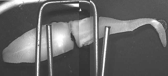
Figure A
Case Answer
This was a case of pentastomiasis. Based on the image and information, the specimen was probably in the family Linguatulidae, although further identification was not possible. Diagnostic features included:
- a general morphology of the worm that ruled-out the possibility of a nematode or trematode.
- pseudosegmention, that ruled-out true proglottids seen in cestodes.
Humans can become infected by ingestion of pentastomid eggs, or eating raw or undercooked meat of herbivorous hosts. Typically these larval worms lodge in various organs, such as the lungs, liver, mesentery, or, rarely, in the eye. In some regions of the world, especially the Middle East, a condition known as halzoun or marrara, results when the larval pentasomtes enter the throat or naso-pharyngeal space in humans, causing nasopharyngitis, with coughing, hemoptysis, vomiting, or nasal discharge. However, there are documented, rare cases where adult worms have been recovered.
Although this case was challenging, it was a good example of the usefulness of telediagnosis. By sharing digital images of suspected parasites, a community of experts is available to provide assistance in difficult cases.
This case was kindly contributed by the Swedish Institute for Infectious Disease Control.
Images presented in the monthly case studies are from specimens submitted for diagnosis or archiving. On rare occasions, clinical histories given may be partly fictitious.
DPDx is an education resource designed for health professionals and laboratory scientists. For an overview including prevention and control visit www.cdc.gov/parasites/.
- Page last reviewed: August 24, 2016
- Page last updated: August 24, 2016
- Content source:
- Global Health – Division of Parasitic Diseases and Malaria
- Notice: Linking to a non-federal site does not constitute an endorsement by HHS, CDC or any of its employees of the sponsors or the information and products presented on the site.
- Maintained By: