


Case #148 - January, 2005
 ShareCompartir
A public health laboratory sent images to DPDx for telediagnosis assistance to confirm their parasite identification of a 160 micrometer long object. The patient was a refugee from Africa. Figure A shows the object in a formalin-ethyl acetate (FEA) concentrated wet mount. What is your diagnosis? Based on what criteria?
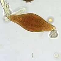
Figure A
Case Answer
This was a case schistosomiasis caused Schistosoma intercalatum, as demonstrated by an egg in an FEA concentrated stool specimen. Diagnostic features seen in the images included:
- the size of the egg, which was consistent with S. intercalatum (140-240 micrometers). It should be noted that the size range overlaps with that of the morpholigcally-similar, S. haematobium (eggs 112-170 micrometers long).
- the shape of the egg, with a prominent equatorial bulge and terminal spine, which is typical for S. intercalatum. Schistosoma haematobium also has a terminal spine, but the egg is generally more evenly rounded.
Eggs of S. intercalatum are typcially found in feces, whereas S. haematobium eggs are usually found in urine. If proper specimen collection techniques are not followed, it is possible for urine to contaminate a fecal specimen, so careful observation of morphologic features is important.
More on: Schistosomiasis
This case was kindly contributed by the Vermont Department of Health Laboratory.
Images presented in the monthly case studies are from specimens submitted for diagnosis or archiving. On rare occasions, clinical histories given may be partly fictitious.
DPDx is an education resource designed for health professionals and laboratory scientists. For an overview including prevention and control visit www.cdc.gov/parasites/.
- Page last reviewed: August 24, 2016
- Page last updated: August 24, 2016
- Content source:
- Global Health – Division of Parasitic Diseases and Malaria
- Notice: Linking to a non-federal site does not constitute an endorsement by HHS, CDC or any of its employees of the sponsors or the information and products presented on the site.
- Maintained By: