Ultrasonography of deep vein thrombosis
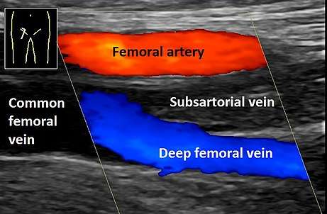
Ultrasonography in suspected deep vein thrombosis focuses primarily on the femoral vein and the popliteal vein, because thrombi in these veins are associated with the greatest risk of harmful pulmonary embolism.
| Ultrasonography of deep vein thrombosis | |
|---|---|
| Medical diagnostics | |
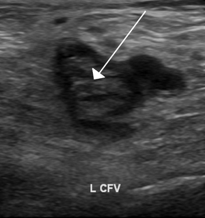 Deep vein thrombosis of the common femoral vein, seen with the probe in a transversal position. | |
| Purpose | focuses primarily on femoral and popliteal vein, |
Medical uses
The risk of deep vein thrombosis can be estimated by Wells score.
Lower limbs venous ultrasonography is also indicated in cases of suspected pulmonary embolism where a CT pulmonary angiogram is negative but a high clinical suspicion of pulmonary embolism remains.[1] It may identify a deep vein thrombosis in up to 50% of people with pulmonary embolism.[1]
Knee or hip replacement are, by themselves, not indications to perform the procedure.[2]
Serial follow-up the ultrasound exam is not necessary after an initially complete, normal study in individuals with DVT symptoms who have suspected pulmonary embolism and nondiagnostic ventilation/perfusion scans .[3]
Technique and findings
Unlike arterial ultrasonography, venous ultrasonography is carried out with the probe in a transversal position, (perpendicular to the vein axis), displaying cross-sections of the veins.[4] All collateral veins are better detected this way, including perforator veins, but of most importance is the detection of venous thrombosis. The most reliable sign of thrombosis (even when a good image and color is present) is the absence of compressibility - A vein cannot be compressed when the blood is in a solid state, as with a thrombus, in the same way that a rubber pipe cannot be compressed if the water inside is frozen.[5] However, if the probe is parallel to the vein axis, when the examiner compresses it, the probe can slide to the right or to the left giving a false negative for thrombosis as the probe has moved away and the vein will not then be evident. Nevertheless, when the examiner needs to show the head thrombus in a printout, the probe will be presented parallel to the vein axis.[4]
Echogenicity
A very recently formed thrombus is not very solid, it will have a low echogenicity, and will be seen as a black area in the gray-scale image and will be hardly visible. When the examiner uses color, the imaging is not much improved.[6] A thrombus may not be evident in the scan. Also a vein lumen may show echoes without the presence of a thrombus. The location of the thrombus and its detail will inform of the seriousness of the condition. In a deep vein thrombosis (DVT), or in a superficial vein thrombosis where the thrombus is floating, an emergency situation will be indicated. If the thrombus is near to the sapheno-femoral junction there will be a high risk of a pulmonary embolism occurring.[7][8]
Compression
The inability to compress the vein is one of the more reliable indications of venous thrombosis. There is a simplified technique called "compression ultrasonography" which can be used for quick DVT diagnosis, mainly for the common femoral vein and the popliteal vein. It is very useful in an emergency situation and is performed just by vein compression using transducer pressure.[9] Compression ultrasonography has both high sensitivity and specificity for detecting DVT in symptomatic patients. Results are not reliable when the patient is symptomless and must be checked carefully. For example, in high risk post-operative patients, mainly after orthopedic surgery where there is already lower limb pain and edema following surgery, thrombi can be localized in the calf veins and often these are not completely occlusive. In this situation a complete examination is mandatory.[10][11][12]
Doppler
Doppler ultrasonography of venous blood flow that correlates with respiration can be diagnostic of the absence of deep vein thrombosis.[13]
See also
- Ultrasonography of chronic venous insufficiency of the legs, mainly targeting superficial veins.
References
- Squizzato, Alessandro; Galli, Luca; Gerdes, Victor E. A. (2015). "Point-of-care ultrasound in the diagnosis of pulmonary embolism". Critical Ultrasound Journal. 7 (1): 7. doi:10.1186/s13089-015-0025-5. ISSN 2036-3176. PMC 4447771. PMID 26034556.
- American Academy of Orthopaedic Surgeons (February 2013), "Five Things Physicians and Patients Should Question", Choosing Wisely: an initiative of the ABIM Foundation, American Academy of Orthopaedic Surgeons, retrieved 19 May 2013, which cites
- Members of 2007 and 2011 AAOS Guideline Development Work Groups on PE/VTED Prophylaxis; Mont, M; Jacobs, J; Lieberman, J; Parvizi, J; Lachiewicz, P; Johanson, N; Watters, W (Apr 18, 2012). "Preventing venous thromboembolic disease in patients undergoing elective total hip and knee arthroplasty". The Journal of Bone and Joint Surgery. American Volume. 94 (8): 673–4. doi:10.2106/JBJS.9408edit. PMC 3326687. PMID 22517384.
- Bendick, Phillip J.; Glover, John L.; Brown, O.William; Ranval, Timothy J. (1996). "Serial duplex ultrasound examinations for deep vein thrombosis in patients with suspected pulmonary embolism". Journal of Vascular Surgery. 24 (5): 732–737. doi:10.1016/S0741-5214(96)70005-8. ISSN 0741-5214.
- Coleridge-Smith, P.; Labropoulos, N.; Partsch, H.; Myers, K.; Nicolaides, A.; Cavezzi, A. (2006). "Duplex Ultrasound Investigation of the Veins in Chronic Venous Disease of the Lower Limbs—UIP Consensus Document. Part I. Basic Principles". European Journal of Vascular and Endovascular Surgery. 31 (1): 83–92. doi:10.1016/j.ejvs.2005.07.019. PMID 16226898.
- Page pp. 89–95 in: Raghavendra BN, Horii SC, Hilton S, Subramanyam BR, Rosen RJ, Lam S; Horii; Hilton; Subramanyam; Rosen; Lam (1986). "Deep venous thrombosis detection by probe compression of veins". J.Ultrasound Med. 5 (2): 89–95. doi:10.7863/jum.1986.5.2.89. PMID 3514943. Archived from the original on 2013-07-25.CS1 maint: multiple names: authors list (link)
- Page 422 in: Dauzat, Michel (1991). Ultrasonographie vasculaire diagnostique: Théorie et pratique [Vascular diagnostic ultrasound: Theory and practice] (in French). Paris: Vigot. pp. 386–437. ISBN 978-2-7114-1104-7.
- Page 1-2 in: "Training in Diagnostic Ultrasound: Essentials, Principles and Standards: Report of a Who Study Group" (PDF). World Health Organization Technical Report Series. 875: i–46, back cover. 1998. ISBN 978-92-4-120875-8. PMID 9659004. Retrieved 2013-02-05.
- Elias, Antoine; Mallard, Luc; Elias, Marie; Alquier, Catherine; Guidolin, François; Gauthier, Bruno; Viard, Alain; Mahouin, Pierre; et al. (2011). "A single complete ultrasound investigation of the venous network for the diagnostic management of patients with a clinically suspected first episode of deep venous thrombosis of the lower limbs". Thrombosis and Haemostasis. 89 (2): 221–7. doi:10.1267/THRO03020221 (inactive 2019-08-20). PMID 12574799.
- Cogo, Alberto; Lensing, AW; Prandoni, P; Hirsh, J (1993). "Distribution of Thrombosis in Patients with Symptomatic Deep Vein Thrombosis: Implications for Simplifying the Diagnostic Process with Compression Ultrasound". Archives of Internal Medicine. 153 (24): 2777–80. doi:10.1001/archinte.1993.00410240085010. PMID 8257253.
- Hollyoak, Maureen; Woodruff, Peter; Muller, Michael; Daunt, Nicholas; Weir, Paula (2001). "Deep venous thrombosis in postoperative vascular surgical patients: A frequent finding without prophylaxis". Journal of Vascular Surgery. 34 (4): 656–60. doi:10.1067/mva.2001.116803. PMID 11668320.
- Elliott, C. Gregory (2000). "The Diagnostic Approach to Deep Venous Thrombosis: Diagnostic Tests for Deep Vein Thrombosis". Seminars in Respiratory and Critical Care Medicine. 21 (6): 495–504. doi:10.1055/s-2000-13187. PMID 16088760.
- Jongbloets, L.M.M.; Koopman, M.M.W.; Büller, H.R.; Ten Cate, J.W.; Lensing, A.W.A. (1994). "Limitations of compression ultrasound for the detection of symptomless postoperative deep vein thrombosis". The Lancet. 343 (8906): 1142–4. doi:10.1016/S0140-6736(94)90240-2. PMID 7910237.
- The Management of Venous Thromboembolic Diseases and the Role of Thrombophilia Testing, NICE Clinical Guidelines, No. 144. National Clinical Guideline Centre (UK). London: Royal College of Physicians (UK); June. Royal College of Physicians (UK). 2012.