Tomotherapy
Tomotherapy is a radiation therapy modality[1][2][3], in which the patient is scanned across a modulated strip-beam, so that only one “slice” (Greek prefix “tomo-”) of the target is exposed at any one time by the linear accelerator (linac) beam. The three components distinctive to this modality are: (1) a collimator pair that defines the length of the strip, (2) a binary multileaf collimator whose leaves open and close during treatment to modulate the strip’s intensity, and (3) a couch that scans the patient across the beam at a fixed speed the during treatment delivery.
| Tomotherapy | |
|---|---|
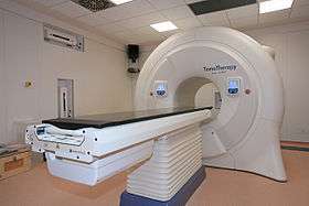 Tomotherapy Hi Art | |
| Other names | Helical tomotherapy |
| Specialty | oncology |
General principles
The treatment field’s length is selectable. In static-jaw delivery, the field length remains constant during a treatment. In dynamic-jaw delivery, the field length changes so that it begins and ends at its minimum setting.

TomoTherapy treatment times vary compared to normal radiation therapy treatment times (tomotherapy treatment times can be as low as 6.5 minutes for common prostate treatment[4]) but do add an additional 2–3 minutes for a daily CT. The daily CT is used to precisely place the radiation beam and allows the operator to modify the treatment should the patient's anatomy change due to weight loss or tumor shrinkage (image-guided radiation therapy).
There are few head to head comparisons of tomotherapy and other IMRT techniques, however there is some evidence that VMAT can provide faster treatment while tomotherapy is better able to spare surrounding healthy tissue while delivering a uniform dose.[5][6][7]
Helical Delivery
In helical tomotherapy, the linac rotates on its gantry at a constant speed while the beam is delivered; so that from the patient’s perspective, the shape traced out by the linac is helical.
While helical tomotherapy can treat very long volumes without a need to abut fields in the longitudinal direction, it does display a distinct artifact due to "thread effect"[8] when treating non-central tumors. Thread effect can be suppressed during planning through good pitch selection.
Fixed-Angle Delivery
Fixed-angle tomotherapy uses multiple tomotherapy beams, each delivered from a separate fixed gantry angle, in which only the couch moves during beam delivery. This is branded as TomoDirect, but has also been called topotherapy[9][10].
The technology enables fixed beam treatments by moving the patient through the machine bore while maintaining specified beam angles.
Clinical Considerations
Lung cancer, head and neck tumors, breast cancer, prostate cancer, stereotactic radiosurgery (SRS) and stereotactic body radiotherapy (SBRT) are some examples of treatments commonly performed using tomotherapy.[11][12][13]
In general, radiation therapy (or radiotherapy) has developed with a strong reliance on homogeneity of dose throughout the tumor. Tomotherapy embodies the sequential delivery of radiation to different parts of the tumor which raises two important issues. First, this method is known as "field matching" and brings with it the possibility of a less-than-perfect match between two adjacent fields with a resultant hot and/or cold spot within the tumor. The second issue is that if the patient or tumor moves during this sequential delivery, then again, a hot or cold spot will result. The first problem is reduced by use of a helical motion, as in spiral computed tomography.[14]
Some research has suggested tomotherapy provides more conformal treatment plans and decreased acute toxicity.[15]
Non-helical static beam techniques such as IMRT and TomoDirect are well suited to whole breast radiation therapy. These treatment modes avoid the low-dose integral splay and long treatment times associated with helical approaches by confining dose delivery to tangential angles. [16] [17] [18]
This risk is accentuated in younger patients with early-stage breast cancer, where cure rates are high and life expectancy is substantial.[18]
Static beam angle approaches aim to maximize the therapeutic ratio by ensuring that the tumor control probability (TCP) significantly outweighs the associated normal tissue complication probability (NTCP).[19][20][21]
History
The tomotherapy technique was developed in the early 1990s at the University of Wisconsin–Madison by Professor Thomas Rockwell Mackie and Paul Reckwerdt.[22] A small megavoltage x-ray source was mounted in a similar fashion to a CT x-ray source, and the geometry provided the opportunity to provide CT images of the body in the treatment setup position. Although original plans were to include kilovoltage CT imaging, current models use megavoltage energies. With this combination, the unit was one of the first devices capable of providing modern image-guided radiation therapy (IGRT).[14]
The first implementation of tomotherapy was the Corvus system developed by Nomos Corporation, with the first patient treated in April, 1994.[23][24] This was the first commercial system for planning and delivering intensity modulated radiation therapy (IMRT). The original system, designed solely for use in the brain, incorporated a rigid skull-based fixation system to prevent patient motion between the delivery of each slice of radiation. But some users [25] eschewed the fixation system and applied the technique to tumors in many different parts of the body.
At this time, the systems manufactured by Accuray (previously TomoTherapy Inc.) are the primary tomotherapy devices in use.
Mobile tomotherapy
Due to their internal shielding and small footprint, TomoTherapy Hi-Art and TomoTherapy TomoHD treatment machines were the only high energy radiotherapy treatment machines used in relocatable radiotherapy treatment suites. Two different types of suites were available: TomoMobile developed by TomoTherapy Inc. which was a moveable truck; and Pioneer, developed by UK-based Oncology Systems Limited. The latter was developed to meet the requirements of UK and European transport law requirements and was a contained unit placed on a concrete pad, delivering radiotherapy treatments in less than five weeks.[26][27]
See also
References
- Mayles, Philip; Nahum, Alan; Rosenwald, Jean-Claude, eds. (2007). Handbook of radiotherapy physics theory and practice. Boca Raton: CRC Press. p. 969. ISBN 9781420012026.
- Colligan, S J; Mills, J (2012). "Beam therapy equipment". In Sibtain, Amen; Morgan, Andrew; MacDougall, Niall (eds.). Radiotherapy in practice : physics for clinical oncology. Oxford: Oxford University Press. doi:10.1093/med/9780199573356.001.0001. ISBN 9780199573356.
- Fenwick, John D.; et al. (October 2006). "Tomotherapy and Other Innovative IMRT Delivery Systems". Seminars in Radiation Oncology. 16 (4): 199–208. doi:10.1016/j.semradonc.2006.04.002. PMID 17010902.
- Piotrowski, T; et al. (June 2014). "Tomotherapy: implications on daily workload and scheduling patients based on three years' institutional experience". Technology in Cancer Research & Treatment. 13 (3): 233–42. doi:10.7785/tcrt.2012.500374. PMID 24066951.
- "VMAT vs. Tomotherapy". Imaging Technology News. 2010-06-02. Retrieved 6 June 2016.
- Rao, M.; et al. (November 2009). "Evaluation of Arc-based Intensity Modulated Radiotherapy for Head and Neck Cancer". International Journal of Radiation Oncology*Biology*Physics. 75 (3): S419. doi:10.1016/j.ijrobp.2009.07.959.
- Oliver, Michael; et al. (15 November 2009). "Comparing planning time, delivery time and plan quality for IMRT, RapidArc and Tomotherapy". Journal of Applied Clinical Medical Physics. 10 (4): 3068. doi:10.1120/jacmp.v10i4.3068. PMC 5720582. PMID 19918236.
- Kissick, M. W.; et al. (May 2005). "The helical tomotherapy thread effect". Med. Phys. 32 (5): 1414–1423. doi:10.1118/1.1896453. PMID 15984692.
- Gonzalez V, Buchholz D, Langen K, et al. Evaluation of two tomotherapy-based techniques for the delivery of whole-breast intensity-modulated radiation therapy. Int J Radiat Oncol Biol Phys 2006; 65: 284–90.
- Gonzalez V, Buchholz D, Langen K, et al. Evaluation of two tomotherapy-based techniques for the delivery of whole-breast intensity-modulated radiation therapy. Int J Radiat Oncol Biol Phys 2006; 65: 284–90.
- Woo, Shiao Y.; et al. (June 1996). "A comparison of intensity modulated conformal therapy with a conventional external beam stereotactic radiosurgery system for the treatment of single and multiple intracranial lesions". International Journal of Radiation Oncology*Biology*Physics. 35 (3): 593–597. doi:10.1016/S0360-3016(96)80023-X. PMID 8655384.
- Cherry, Pam; Duxbury, Angela, eds. (2009). Practical radiotherapy : physics and equipment (2nd ed.). Chichester: Wiley-Blackwell. p. 210. ISBN 9781405184267.
- Peñagarícano, José A; et al. (2006). "Dosimetric comparison of Helical Tomotherapy and Gamma Knife Stereotactic Radiosurgery for single brain metastasis". Radiation Oncology. 1 (1): 26. doi:10.1186/1748-717X-1-26. PMC 1557668. PMID 16887031.
- Mackie, T R (7 July 2006). "History of tomotherapy". Physics in Medicine and Biology. 51 (13): R427–R453. doi:10.1088/0031-9155/51/13/R24. PMID 16790916.
- Yu, Mina; et al. (2013). "A comparison of dosimetric parameters between tomotherapy and three-dimensional conformal radiotherapy in rectal cancer". Radiation Oncology. 8 (1): 181. doi:10.1186/1748-717X-8-181. PMC 3721992. PMID 23866263.
- Squires M, Hu Y, Byrne M, et al. Static beam tomotherapy as an optimisation method in whole breast radiation therapy (WBRT). J Med Radiat Sci, http://onlinelibrary.wiley.com/doi/10.1002/jmrs.232/abstract
- Goddu SM, Chaudhari S, Mamalui-Hunter M, et al. Helical tomotherapy planning for left-sided breast cancer patients with positive lymph nodes: Comparison to conventional multiport breast technique. Int J Radiat Oncol Biol Phys 2009; 73: 1243–51.
- Stovall M, Smith SA, Langholz BM, et al. Dose to the contralateral breast from radiotherapy and risk of second primary breast cancer in the WECARE study. Int J Radiat Oncol Biol Phys 2008; 72: 1021–30.
- Franco P, Catuzzo P, Cante D, et al. TomoDirect: An efficient means to deliver radiation at static angles with tomotherapy. Tumori 2011; 97: 498–502.
- Franco P, Ricardi U. Tomo Direct to deliver static angle tomotherapy treatments. J Nucl Med Radiat Ther 2012; 3:5.
- Murai T, Shibamoto Y, Manabe Y, et al. Intensity modulated radiation therapy using ports of Tomotherapy (TomoDirect): Comparison with the TomoHelical mode. J Radiat Oncol 2013; 8: 68.
- Holmes, Timothy W.; et al. (June 2008). "Stereotactic Image-Guided Intensity Modulated Radiotherapy Using the HI-ART II Helical Tomotherapy System". Medical Dosimetry. 33 (2): 135–148. doi:10.1016/j.meddos.2008.02.006. PMID 18456165.
- Mackie, T. Rockwell; et al. (January 1999). "Tomotherapy". Seminars in Radiation Oncology. 9 (1): 108–117. doi:10.1016/S1053-4296(99)80058-7. PMID 10196402.
- Woo, Shiao Y.; et al. (June 1996). "A comparison of intensity modulated conformal therapy with a conventional external beam stereotactic radiosurgery system for the treatment of single and multiple intracranial lesions". International Journal of Radiation Oncology*Biology*Physics. 35 (3): 593–597. doi:10.1016/S0360-3016(96)80023-X. PMID 8655384.
- Squires M, Hu Y, Byrne M, et al. Static beam tomotherapy as an optimisation method in whole breast radiation therapy (WBRT). J Med Radiat Sci, Forthcoming 2017.
- "Radiation Therapy on the Road". Imaging Technology News. 2010-04-19. Retrieved 6 June 2016.
- "OSL launches Pioneer relocatable radiotherapy suite". medicalphysicsweb.org. Retrieved 6 June 2016.
Nuclear technology | |||||||||||||||||||||||||||||||||||||||||||||||||||||||
|---|---|---|---|---|---|---|---|---|---|---|---|---|---|---|---|---|---|---|---|---|---|---|---|---|---|---|---|---|---|---|---|---|---|---|---|---|---|---|---|---|---|---|---|---|---|---|---|---|---|---|---|---|---|---|---|
| |||||||||||||||||||||||||||||||||||||||||||||||||||||||
| |||||||||||||||||||||||||||||||||||||||||||||||||||||||
| |||||||||||||||||||||||||||||||||||||||||||||||||||||||