Psammoma body
A psammoma body is a round collection of calcium, seen microscopically. The term is derived from the Greek word ψάμμος (psámmos), meaning "sand".
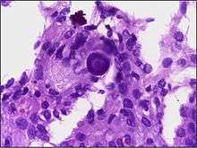
Micrograph of psammoma body in the centre of the field in a meningioma of brain. H&E stain.
Cause
Psammoma bodies are associated with the papillary (nipple-like) histomorphology and are thought to arise from,
- Infarction and calcification of papillae tips.
- Calcification of intralymphatic tumor thrombi.[1]
Association with lesions
Psammoma bodies are commonly seen in certain tumors such as:
- Papillary thyroid carcinoma
- Papillary renal cell carcinoma
- Ovarian papillary serous cystadenoma and cystadenocarcinoma[2]
- Endometrial adenocarcinomas (Papillary serous carcinoma ~3%-4%)
- Meningiomas, in the central nervous system[3]
- Peritoneal and Pleural Mesothelioma
- Somatostatinoma (pancreas)[4]
- Prolactinoma of the pituitary [5]
- Glucagonoma
- Micropapillary subtype of Lung Adenocarcinoma[6]
Benign lesions
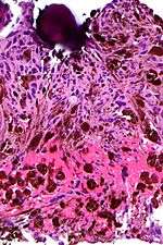
Micrograph of a psammomatous melanotic schwannoma with a psammoma body, as may be seen in Carney complex. H&E stain.
Psammoma bodies may be seen in:
- Endosalpingiosis[7]
- Psammomatous melanotic schwannoma
- Melanocytic nevus[8]
Appearance
Psammoma bodies usually have a laminar appearance, are circular, acellular and basophilic.
References
- Johannessen JV, Sobrinho-Simões M (September 1980). "The origin and significance of thyroid psammoma bodies". Lab. Invest. 43 (3): 287–96. PMID 7401638.
- Ovarian papillary serous cystadenocarcinoma at WebPath, The Internet Pathology Laboratory for Medical Education at Mercer University School of Medicine. Retrieved July 2011
- http://spinwarp.ucsd.edu/neuroweb/Text/br-300b.htm
- "Pancreatic Endocrine Tumors: Radiologic-Clinicopathologic Correlation". RadioGraphics. 30: 1445–1464. doi:10.1148/rg.306105523.
- Robbin's Pathology, Eight Ed
- 3. Expansion of the concept of micropapillary adenocarcinoma to include a newly recognized filigree pattern as well as the classical pattern based on 1468 Stage I lung adenocarcinomas. Emoto K, Eguchi T et al. J Thorac Oncol. 2019 Jul 25. link
- Hallman KB, Nahhas WA, Connelly PJ (September 1991). "Endosalpingiosis as a source of psammoma bodies in a Papanicolaou smear. A case report". J Reprod Med. 36 (9): 675–8. PMID 1774734.
- Rapini, Ronald. Practical Dermatopathology. Elsevier Mosby, 2005, p. 10.
This article is issued from
Wikipedia.
The text is licensed under Creative
Commons - Attribution - Sharealike.
Additional terms may apply for the media files.