Phytoplasma
Phytoplasmas are obligate bacterial parasites of plant phloem tissue and of the insect vectors that are involved in their plant-to-plant transmission. Phytoplasmas were discovered in 1967 by Japanese scientists who termed them mycoplasma-like organisms.[2] Since their discovery, phytoplasmas have resisted all attempts at in vitro culture in any cell-free medium; routine cultivation in an artificial medium thus remains a major challenge. Although phytoplasmas have recently been reported to be grown in a specific artificial medium, experimental repetition has yet to be reported.[3] Phytoplasmas are characterized by the lack of a cell wall, a pleiomorphic or filamentous shape, a diameter normally less than 1 μm, and a very small genome.
| Phytoplasma | |
|---|---|
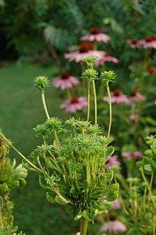 | |
| Phyllody induced by phytoplasma infection on a coneflower (Echinacea purpurea) | |
| Scientific classification | |
| Domain: | Bacteria |
| Phylum: | Tenericutes |
| Class: | Mollicutes |
| Order: | Acholeplasmatales |
| Family: | Acholeplasmataceae |
| Genus: | "Candidatus Phytoplasma" |
| Species | |
| |
Phytoplasmas are pathogens of agriculturally important plants, including coconut, sugarcane, and sandalwood, in which they cause a wide variety of symptoms ranging from mild yellowing to death. Phytoplasmas are most prevalent in tropical and subtropical regions. They are transmitted from plant to plant by vectors (normally sap-sucking insects such as leafhoppers) in which they both survive and replicate.
History
References to diseases now known to be caused by phytoplasmas can be found as far back as 1603 (mulberry dwarf disease in Japan.)[4] Such diseases were originally thought to be caused by viruses, which, like phytoplasmas, require insect vectors, and cannot be cultured. Viral and phytoplasmic infections share some symptoms.[5] In 1967, phytoplasmas were discovered in ultrathin sections of plant phloem tissue and were termed mycoplasma-like organisms due to their physiological resemblance[2] The organisms were renamed phytoplasmas in 1994, at the 10th Congress of the International Organization for Mycoplasmology.[5]
Morphology
Phytoplasmas are Mollicutes, which are bound by a triple-layered membrane, rather than a cell wall.[6] The phytoplasma cell membranes studied to date usually contain a single immunodominant protein of unknown function that constitutes most of the protein in the membrane.[7] A typical phytoplasma is pleiomorphic or filamentous in shape and is less than 1 μm in diameter. Like other prokaryotes, phytoplasmic DNA is distributed throughout the cytoplasm, instead of being concentrated in a nucleus.
Symptoms
Phytoplasmas can infect and cause various symptoms in more than 700 plant species. One characteristic symptom is abnormal floral organ development including phyllody, (i.e., the production of leaf-like structures in place of flowers) and virescence (i.e., the development of green flowers attributable to a loss of pigment by petal cells).[8] Phytoplasma-harboring flowering plants may nevertheless be sterile. The expression of genes involved in maintaining the apical meristem or in the development of floral organs is altered in the morphologically affected floral organs of phytoplasma-infected plants.[9][10]
A phytoplasma infection often triggers leaf yellowing, probably due to the presence of phytoplasma cells in phloem, which can affect phloem function and carbohydrate transport,[11] inhibit chlorophyll biosynthesis, and trigger chlorophyll breakdown.[6] These symptoms may be attributable to stress caused by the infection rather than a specific pathogenetic process.
Many phytoplasma-infected plants develop a bushy or "witch's broom" appearance due to changes in their normal growth patterns. Most plants exhibit apical dominance but infection can trigger the proliferation of axillary (side) shoots and a reduction in internode size.[8] Such symptoms are actually useful in the commercial production of poinsettias. Infection triggers more axillary shoot production; the poinsettia plants thus produce more than a single flower.[12]
Effector (virulence) proteins
Many plant pathogens produce virulence factors (i.e., effectors) that modulate or interfere with normal host processes to the benefit of the pathogens. In 2009, a secreted protein, termed “tengu-su inducer” (TENGU; C0H5W6), was identified from a phytoplasma causing yellowing of onions; this was the first phytoplasmal virulence factor to be described. TENGU induces characteristic symptoms (termed “tengu-su”), including witches’ broom and dwarfism.[13] Transgenic expression of TENGU in Arabidopsis plants induced sterility in male and female flowers.[14] TENGU contains a signal peptide at its N-terminus; after cleavage, the mature protein is only 38 amino acids in length.[13] Although phytoplasmas are restricted to phloem, TENGU is transported from phloem to other cells, including those of the apical and axillary meristems.[13] TENGU was suggested to inhibit both auxin- and jasmonic acid-related pathways, thereby affecting plant development.[13][14] Surprisingly, the N-terminal 11 amino acid region of the mature protein triggers symptom development in Nicotiana benthamiana plants.[15] TENGU undergoes proteolytic processing by a plant serine protease in vivo, suggesting that the N-terminal peptide (i.e., the 11 amino acid fragment) alone induces the observed symptoms. TENGU homologs have been identified in AY-group phytoplasmas. All such homologs undergo processing and can induce symptoms, suggesting that the symptom-inducing mechanism is conserved among TENGU homologs.[15]
In 2009, 56 genes for secreted proteins were identified in the genome of Aster Yellows phytoplasma strain Witches Broom (AY-WB); these were named secreted AY-WB proteins (SAPs) and considered effectors.[16] Also in 2009, effector SAP11 was shown to target plant cell nuclei and unload from phloem cells in AY-WB-infected plants.[16] SAP11 was found to induce stem proliferations and changes of leaf shapes of plants; the stem proliferations induced by SAP11 resemble witch's broom symptoms of AY-WB-infected plants.[17] In addition, it was demonstrated that SAP11 interacts with and destabilizes plant class II TCP protein domain transcription factors that leads to shoot proliferations and leaf shape changes.[17][18] In addition to regulation of plant development, TCPs also control the expression of lipoxygenase genes required for jasmonate biosynthesis.[19][20] Jasmonate levels are decreased in phytoplasma-infected Arabidopsis plants and plants that transgenically express the AY-WB SAP11 effector. The downregulation of jasmonate production is beneficial to phytoplasmas because jasmonate is involved in plant defenses against herbivorous insects such as leafhoppers.[17][21] Leafhoppers lay increased numbers of eggs on AY-WB-infected plants, at least in part because of SAP11 production. For example, the leafhopper Macrosteles quadrilineatus laid 30% more eggs on plants that expressing SAP11 transgenically than control plants, and 60% more eggs on plants infected with AY-WB.[22] Phytoplasmas cannot survive in the external environment and are dependent upon insects such as leafhoppers for transmission to new (healthy) plants. Thus, by compromising jasmonate production, SAP11 'encourages' leafhoppers to lay more eggs on phytoplasma-infected plants, thereby ensuring that newly hatched leafhopper nymphs feed upon infected plants to become phytoplasma vectors. SAP11 effectors are identified in a number of divergent phytoplasmas and these effectors also interact with TCPs and modulate plant defenses.[23][24][25][26] SAP11 is the first phytoplasma virulence protein for which plant targets and effector functions (i.e. evidence of benefit for the pathogen) were identified. TCPs were found to be targeted by a number of other pathogen effectors.[27][28]
The AY-WB phytoplasma effector SAP54 was shown to induce virescence and phyllody when expressed in plants and homologs of this effector were found in at least three other phytoplasmas.[29] Two SAP54 homologs, PHYL1 of the onion yellows phytoplasma and PHYL1PnWB of the peanut witches’ broom phytoplasma, also induce phyllody-like floral abnormalities.[30][31] These results suggest that PHYL1, SAP54, and their homologs form a phyllody-inducing gene family, the members of which are termed phyllogens.[30] MADS-box transcription factors (MTFs) of the ABCE model play critical roles in floral organ development in Arabidopsis. Phyllogens interact directly with class A and class E MTFs, inducing protein degradation in a ubiquitin/proteasome-dependent manner that, at least for SAP54, is dependent on interactions with the proteasome shuttle factor RAD23.[30][32][33] Interestingly, RAD23 mutants do not show phyllody when infected with phytoplasma indicating that RAD23 proteins are susceptibility factors; i.e. phytoplasmas and SAP54 require these plant proteins to induce phyllody symptoms.[34] The accumulation of mRNAs encoding class B MTFs, the transcription of which is positively regulated by class A and class E MTFs, is drastically decreased in Arabidopsis constitutively expressing PHYL1.[30] Phyllogens induce abnormal floral organ development by inhibiting the functions of these MTFs. RAD23 proteins are also required for promoting leafhopper vector egg laying on plants that express SAP54 and are infected with AY-WB phytoplasma.[34][35]
Transmission
Movement between plants
Phytoplasmas are spread principally by insects of the families Cicadellidae (leafhoppers), Fulgoridae (planthoppers), and Psyllidae (jumping plant lice) ,[36] which feed on the phloem of infected plants, ingesting phytoplasmas and transmitting them to the next plant on which they feed. Thus, the host range of phytoplasmas is strongly dependent upon that of the insect vector. Phytoplasmas contain a major antigenic protein constituting most of the cell surface protein. This protein associates with insect microfilament complexes and is believed to control insect-phytoplasma interactions.[37] Phytoplasmas can overwinter in insect vectors or perennial plants. Phytoplasmas can have varying effects on their insect hosts; examples of both reduced and increased fitness have been noted.[38]
Phytoplasmas enter the insect body through the stylet, pass through the intestine, and then move to the hemolymph[38] and colonize the salivary glands: the entire process can take up to 3 weeks.[38] Once established in an insect host, phytoplasmas are found in most major organs. The time between ingestion by the insect and attainment of an infectious titer in the salivary glands is termed the latency period.[38]
Phytoplasmas can also be spread via dodders (Cuscuta)[39] or by vegetative propagation such as the grafting of infected plant tissue onto a healthy plant.
Detection and diagnosis
Before the molecular era, the diagnosis of phytoplasma-caused diseases was difficult because the organisms could not be cultured. Thus, classical diagnostic techniques, including symptom observation were used. Ultrathin sections of phloem tissue from plants with suspected phytoplasma-infections were also studied.[2] The empirical use of antibiotics such as tetracycline was additionally employed.
Molecular diagnostic techniques for phytoplasma detection began to emerge in the 1980s and included enzyme-linked immunosorbent assay (ELISA)-based methods. In the early 1990s, polymerase chain reaction (PCR)-based techniques were developed: these are far more sensitive than ELISAs, and restriction fragment length polymorphism (RFLP) analysis allowed the accurate identification of various phytoplasma strains and species.[41]
More recent techniques allow infection levels to be assessed. Both quantitative PCR and bioimaging can effectively quantify phytoplasma titers within plant.[40] In addition, loop-mediated isothermal amplification (a sensitive, simple, and rapid diagnostic method) is now available as a commercial kit allowing all known phytoplasma species to be detected in about 1 h, including the DNA extraction step.
Control
Phytoplasmas are normally controlled by the breeding and planting of disease-resistant crop varieties (perhaps the most economically viable option) and by the control of insect vectors.[8]
Tissue culture can be used to produce healthy clones of phytoplasma-infected plants. Cryotherapy (i.e., the freezing of plant samples in liquid nitrogen) prior to tissue culture increases the probability of producing healthy plants in this manner.[42]
Plantibodies targeting phytoplasmas have also been developed.[43]
Tetracyclines are bacteriostatic to phytoplasmas.[44] However, disease symptoms reappear in the absence of continuous antibiotic application. Thus, tetracycline is not a viable agricultural control agent, but it is used to protect ornamental coconut trees.[45]
Genetics
The genomes of four phytoplasmas have been sequenced: "onion yellows",[46] "aster yellows witches' broom" (Candidatus [Ca] Phytoplasma asteris),[47] Ca. Phytoplasma australiense,[48] and Ca. Phytoplasma Mali.[49] Phytoplasmas have very small genomes, with extremely small amount of G and C nucleotides (sometimes as little as 23%, which is thought to be the lower threshold for a viable genome).[50] In fact, the Bermuda grass white-leaf phytoplasma has a genome size of only 530 kb, one of the smallest known genomes of all living organisms.[51] The larger phytoplasma genomes are around 1350 kb in size. The small genome size of phytoplasma is attributable to reductive evolution from Bacillus/Clostridium ancestors. Phytoplasmas have lost ≥75% of their original genes, and can thus no longer survive outside of insects or plant phloem. Some phytoplasmas contain extrachromosomal DNA such as plasmids.[52]
Despite their small genomes, many predicted phytoplasma genes are present in multiple copies. Phytoplasmas lack many genes encoding standard metabolic functions and have no functioning homologous recombination pathway, but they do have a sec transport pathway.[47] Many phytoplasmas contain two rRNA operons. Unlike other Mollicutes, the triplet code of UGA is used as a stop codon in phytoplasmas.[53]
Phytoplasma genomes contain large numbers of transposons and insertion sequences and also contain a unique family of repetitive extragenic palindromes termed PhREPS for which no role is known. However, it is theorized that the stem-loop structures in PhREPS play a role in transcription termination or genome stability.[54]
Taxonomy
Phytoplasmas belong to the monotypic order Acholeplasmatales.[8] In 1992, the Subcommittee on the Taxonomy of Mollicutes proposed the use of "Phytoplasma" rather than "mycoplasma-like organisms" "for reference to the phytopathogenic mollicutes".[55] In 2004, the generic name phytoplasma was adopted and is currently of Candidatus (Ca.) status[56] (used for bacteria that cannot be cultured).[57] Phytoplasma taxonomy is complicated because the organisms cannot be cultured; methods normally used to classify prokaryotes are thus not available.[8] Phytoplasma taxonomic groups are based on differences in fragment sizes produced by restriction digests of 16S ribosomal RNA gene sequences (RFLPs) or by comparisons of DNA sequences from 16s/23s spacer regions.[58] The actual number of taxonomic groups remains unclear; recent work on computer-simulated restriction digests of the 16Sr gene suggested up to 28 groups,[59] whereas others have proposed fewer groups, but more subgroups. Each group includes at least one Ca. Phytoplasma species, characterized by distinctive biological, phytopathological, and genetic properties.
Gallery
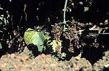 A grape vine with "bois noir" phytoplasma disease
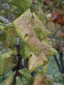 A grape vine with "flavescence dorée" phytoplasma disease
 Coconut palms dying of lethal yellowing disease
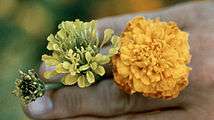 Symptoms of aster yellows on marigold
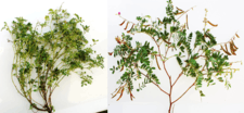 Tephrosia purpurea witch's broom[60]
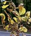 Symptoms of elm phloem necrosis phytoplasma
 Brinjal Little leaf phytoplasma
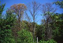 Trees dying of ash yellows phytoplasma
 Parthenium hysterophorus showing symptoms of witch's broom
 Phyllody caused by phytoplasma infection on Cosmos spp.
 Little leaf disease of Cleome viscosa
 Symptoms of sweet potato little leaf phytoplasma on Catharanthus roseus
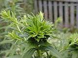 Phyllody of goldenrod
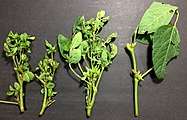 Soybean Phytoplasma
 A flower of China Aster showing phyllody symptoms
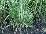 Sugarcane grassy shoot disease[61]
 A palm tree dying of lethal yellowing phytoplasma
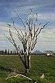 A cabbage tree killed by Phytoplasma australiense
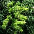 Witch's Broom disease of bamboo (Dendrocalamus strictus)
See also
- Aster yellows
- Elm yellows
- Grapevine yellows
- Mollicutes
- Mycoplasma
- Polymerase chain reaction
- Sugarcane grassy shoot disease
References
- Marcone C, Gibb KS, Streten C, Schneider B (2004). "'Candidatus Phytoplasma spartii','Candidatus Phytoplasma rhamni' and 'Candidatus Phytoplasma allocasuarinae', respectively associated with spartium witches'-broom, buckthorn witches'-broom and allocasuarina yellows diseases". International Journal of Systematic and Evolutionary Microbiology. 54 (4): 1025–1029. doi:10.1099/ijs.0.02838-0. PMID 15280265.
- Doi Y, Teranaka M, Yora K, Asuyama H (1967). "Mycoplasma or PLT-group-like organisms found in the phloem elements of plants infected with mulberry dwarf, potato witches' broom, aster yellows or paulownia witches' broom". Annals of the Phytopathological Society of Japan. 33 (4): 259–266. doi:10.3186/jjphytopath.33.259.
- Contaldo, N.; Bertaccini, A.; Paltrinieri, S.; Windsor, H.M.; Windsor, G.D. (2012). "Axenic culture of a plant pathogenic Phytoplasma". Phytopathologia Mediterranea. 244: 607–617. doi:10.14601/Phytopathol_Mediterr-11773 (inactive 2019-12-06).
- Okuda, S (1972). "Occurrence of diseases caused by mycoplasma-like organisms in Japan". Plant Protection. 26: 180–183.
- Hogenhout, SA; Oshima K; Ammar E-D; Kakizawa S; Kingdom HN; Namba S (2008). "Phytoplasmas: bacteria that manipulate plants and insects". Molecular Plant Pathology. 9 (4): 403–423. doi:10.1111/j.1364-3703.2008.00472.x. PMC 6640453. PMID 18705857. Archived from the original on 2012-10-01. Retrieved 2008-07-04.
- Bertamini, M; Grando M. S; Nedunchezhian N (2004). "Effects of Phytoplasma Infection on Pigments, Chlorophyll-Protein Complex and Photosynthetic Activities in Field Grown Apple Leaves". Biologia Plantarum. 47 (2): 237–242. doi:10.1006/pmpp.2003.0450.
- Berg, M; Davis DL; Clark MF; Vetten HJ; Maier G; Marcone C; Seemuller E (1999). "Isolation of the gene encoding an immunodominant membrane protein of the apple proliferation phytoplasma, and expression and characterization of the gene product". Microbiology. 145 (8): 1939–1943. doi:10.1099/13500872-145-8-1937. PMID 10463160.
- Lee, IM; Davis RE; Gundersen-Rindal DE (2000). "Phytoplasma: Phytopathogenic Mollicutes". Annual Review of Microbiology (Submitted manuscript). 54: 221–255. doi:10.1146/annurev.micro.54.1.221. PMID 11018129.
- Pracros, P; Renaudin J; Eveillard S; Mouras A; Hernould M (2006). "Tomato Flower Abnormalities Induced by Stolbur Phytoplasma Infection Are Associated with Changes of Expression of Floral Development Genes". Molecular Plant-Microbe Interactions. 19 (1): 62–68. doi:10.1094/MPMI-19-0062. PMID 16404954.
- Himeno, M; Neriya Y; Minato N; Miura C; Sugawara K; Ishii Y; Yamaji Y; Kakizawa S; Oshima K; Namba S (2011). "Unique morphological changes in plant pathogenic phytoplasma-infected petunia flowers are related to transcriptional regulation of floral homeotic genes in an organ-specific manner". Plant Journal. 67 (6): 971–979. doi:10.1111/j.1365-313X.2011.04650.x. PMID 21605209.
- Muast BE, Espadas F, Talavera C, Aguilar M, Santamaría JM, Oropeza C (2003). "Changes in carbohydrate metabolism in coconut palms infected with the lethal yellowing phytoplasma". Phytopathology. 93 (8): 976–981. doi:10.1094/PHYTO.2003.93.8.976. PMID 18943864.
- Lee, IM; Klopmeyer M; Bartoszyk IM; Gundersen-Rindal DE; Chou TS; Thomson KL; Eisenreich R (1997). "Phytoplasma induced free-branching in commercial poinsettia cultivars". Nature Biotechnology. 15 (2): 178–182. doi:10.1038/nbt0297-178. PMID 9035146.
- Hoshi, A; Oshima K; Kakizawa S; Ishii Y; Ozeki J; Hashimoto M; Komatsu K; Kagiwada S; Yamaji Y; Namba S (2009). "A unique virulence factor for proliferation and dwarfism in plants identified from a phytopathogenic bacterium". Proceedings of the National Academy of Sciences. 106 (15): 6416–6421. Bibcode:2009PNAS..106.6416H. doi:10.1073/pnas.0813038106. PMC 2669400. PMID 19329488.
- Minato, N; Himeno M; Hoshi A; Maejima K; Komatsu K; Takebayashi Y; Kasahara H; Yusa A; Yamaji Y; Oshima K; Kamiya Y; Namba S (2014). "The phytoplasmal virulence factor TENGU causes plant sterility by downregulating of the jasmonic acid and auxin pathways". Scientific Reports. 4: 7399. Bibcode:2014NatSR...4E7399M. doi:10.1038/srep07399. PMC 4261181. PMID 25492247.
- Sugawara, K; Honma Y; Komatsu K; Himeno M; Oshima K; Namba S (2013). "The alteration of plant morphology by small peptides released from the proteolytic processing of the bacterial peptide TENGU". Plant Physiology. 162 (4): 2004–2015. doi:10.1104/pp.113.218586. PMC 3729778. PMID 23784461.
- Bai, X; Correa, VR; Toruño, TY; Ammar, el-D; Kamoun, S; Hogenhout, SA (January 2009). "AY-WB phytoplasma secretes a protein that targets plant cell nuclei". Molecular Plant-Microbe Interactions. 22 (1): 18–30. doi:10.1094/MPMI-22-1-0018. PMID 19061399.
- Sugio, A; Kingdom, HN; MacLean, AM; Grieve, VM; Hogenhout, SA (29 November 2011). "Phytoplasma protein effector SAP11 enhances insect vector reproduction by manipulating plant development and defense hormone biosynthesis". Proceedings of the National Academy of Sciences of the United States of America. 108 (48): E1254–63. doi:10.1073/pnas.1105664108. PMC 3228479. PMID 22065743.
- Sugio, A; MacLean, AM; Hogenhout, SA (May 2014). "The small phytoplasma virulence effector SAP11 contains distinct domains required for nuclear targeting and CIN-TCP binding and destabilization". The New Phytologist. 202 (3): 838–48. doi:10.1111/nph.12721. PMC 4235307. PMID 24552625.
- Schommer, C; Debernardi, JM; Bresso, EG; Rodriguez, RE; Palatnik, JF (October 2014). "Repression of cell proliferation by miR319-regulated TCP4". Molecular Plant. 7 (10): 1533–44. doi:10.1093/mp/ssu084. PMID 25053833.
- Danisman, S; van der Wal, F; Dhondt, S; Waites, R; de Folter, S; Bimbo, A; van Dijk, AD; Muino, JM; Cutri, L; Dornelas, MC; Angenent, GC; Immink, RG (August 2012). "Arabidopsis class I and class II TCP transcription factors regulate jasmonic acid metabolism and leaf development antagonistically". Plant Physiology. 159 (4): 1511–23. doi:10.1104/pp.112.200303. PMC 3425195. PMID 22718775.
- Kallenbach, M; Bonaventure, G; Gilardoni, PA; Wissgott, A; Baldwin, IT (12 June 2012). "Empoasca leafhoppers attack wild tobacco plants in a jasmonate-dependent manner and identify jasmonate mutants in natural populations". Proceedings of the National Academy of Sciences of the United States of America. 109 (24): E1548–57. Bibcode:2012PNAS..109E1548K. doi:10.1073/pnas.1200363109. PMC 3386116. PMID 22615404.
- Sugio, A; Kingdom HN; MacLean AM; Grieve VM; Hogenhout SA (2011). "Phytoplasma protein effector SAP11 enhances insect vector reproduction by manipulating plant development and defense hormone biosynthesis". Proceedings of the National Academy of Sciences. 108 (48): E1254–E1263. doi:10.1073/pnas.1105664108. PMC 3228479. PMID 22065743.
- Janik, K; Mithöfer, A; Raffeiner, M; Stellmach, H; Hause, B; Schlink, K (April 2017). "An effector of apple proliferation phytoplasma targets TCP transcription factors-a generalized virulence strategy of phytoplasma?". Molecular Plant Pathology. 18 (3): 435–442. doi:10.1111/mpp.12409. PMC 6638208. PMID 27037957.
- Tan, CM; Li, CH; Tsao, NW; Su, LW; Lu, YT; Chang, SH; Lin, YY; Liou, JC; Hsieh, LC; Yu, JZ; Sheue, CR; Wang, SY; Lee, CF; Yang, JY (July 2016). "Phytoplasma SAP11 alters 3-isobutyl-2-methoxypyrazine biosynthesis in Nicotiana benthamiana by suppressing NbOMT1". Journal of Experimental Botany. 67 (14): 4415–25. doi:10.1093/jxb/erw225. PMC 5301940. PMID 27279277.
- Wang, N; Yang, H; Yin, Z; Liu, W; Sun, L; Wu, Y (December 2018). "Phytoplasma effector SWP1 induces witches' broom symptom by destabilizing the TCP transcription factor BRANCHED1". Molecular Plant Pathology. 19 (12): 2623–2634. doi:10.1111/mpp.12733. PMC 6638060. PMID 30047227.
- Chang, SH; Tan, CM; Wu, CT; Lin, TH; Jiang, SY; Liu, RC; Tsai, MC; Su, LW; Yang, JY (26 November 2018). "Alterations of plant architecture and phase transition by the phytoplasma virulence factor SAP11". Journal of Experimental Botany. 69 (22): 5389–5401. doi:10.1093/jxb/ery318. PMC 6255702. PMID 30165491.
- Mukhtar, MS; Carvunis, AR; Dreze, M; Epple, P; Steinbrenner, J; Moore, J; Tasan, M; Galli, M; Hao, T; Nishimura, MT; Pevzner, SJ; Donovan, SE; Ghamsari, L; Santhanam, B; Romero, V; Poulin, MM; Gebreab, F; Gutierrez, BJ; Tam, S; Monachello, D; Boxem, M; Harbort, CJ; McDonald, N; Gai, L; Chen, H; He, Y; European Union Effectoromics, Consortium.; Vandenhaute, J; Roth, FP; Hill, DE; Ecker, JR; Vidal, M; Beynon, J; Braun, P; Dangl, JL (29 July 2011). "Independently evolved virulence effectors converge onto hubs in a plant immune system network". Science. 333 (6042): 596–601. Bibcode:2011Sci...333..596M. doi:10.1126/science.1203659. PMC 3170753. PMID 21798943.
- Yang, L; Teixeira, PJ; Biswas, S; Finkel, OM; He, Y; Salas-Gonzalez, I; English, ME; Epple, P; Mieczkowski, P; Dangl, JL (8 February 2017). "Pseudomonas syringae Type III Effector HopBB1 Promotes Host Transcriptional Repressor Degradation to Regulate Phytohormone Responses and Virulence". Cell Host & Microbe. 21 (2): 156–168. doi:10.1016/j.chom.2017.01.003. PMC 5314207. PMID 28132837.
- MacLean, A. M.; Sugio, A.; Makarova, O. V.; Findlay, K. C.; Grieve, V. M.; Toth, R.; Nicolaisen, M.; Hogenhout, S. A. (2011). "Phytoplasma effector SAP54 induces indeterminate leaf-like flower development in Arabidopsis plants". Plant Physiology. 157 (2): 831–841. doi:10.1104/pp.111.181586. PMC 3192582. PMID 21849514.
- Maejima, K; Iwai R; Himeno M; Komatsu K; Kitazawa Y; Fujita N; Ishikawa K; Fukuoka M; Minato N; Yamaji Y; Oshima K; Namba S (2014). "Recognition of floral homeotic MADS-domain transcription factors by a phytoplasmal effector, phyllogen, induces phyllody". Plant Journal. 78 (4): 541–554. doi:10.1111/tpj.12495. PMC 4282529. PMID 24597566.
- Yang, CY; Huang YH; Lin CP; Lin YY; Hsu HC; Wang CN; Liu LY; Shen B; Lin SS (2015). "MiR396-targeted SHORT VEGETATIVE PHASE is required to repress flowering and is related to the development of abnormal flower symptoms by the PHYL1 effector". Plant Physiology. 168 (4): 1702–1716. doi:10.1104/pp.15.00307. PMC 4528741. PMID 26103992.
- MacLean, AM; Orlovskis Z; Kowitwanich K; Zdziarska AM; Angenent GC; Immink RG; Hogenhout SA (2014). "Phytoplasma effector SAP54 hijacks plant reproduction by degrading MADS-box proteins and promotes insect colonization in a RAD23-dependent manner". PLoS Biology. 12 (4): 831–841. doi:10.1104/pp.111.181586. PMC 3192582. PMID 21849514.
- Maejima, K; Kitazawa Y; Tomomitsu T; Yusa A; Neriya Y; Himeno M; Yamaji Y; Oshima K; Namba S (2015). "Degradation of class E MADS-domain transcription factors in Arabidopsis by a phytoplasmal effector, phyllogen". Plant Signaling & Behavior. 10 (8): e1042635. doi:10.1080/15592324.2015.1042635. PMC 4623417. PMID 26179462.
- MacLean, AM; Sugio, A; Makarova, OV; Findlay, KC; Grieve, VM; Tóth, R; Nicolaisen, M; Hogenhout, SA (October 2011). "Phytoplasma effector SAP54 induces indeterminate leaf-like flower development in Arabidopsis plants". Plant Physiology. 157 (2): 831–41. doi:10.1104/pp.111.181586. PMID 21849514.
- Orlovskis, Z; Hogenhout, SA (2016). "A Bacterial Parasite Effector Mediates Insect Vector Attraction in Host Plants Independently of Developmental Changes". Frontiers in Plant Science. 7: 885. doi:10.3389/fpls.2016.00885. PMC 4917533. PMID 27446117.
- Weintraub, Phyllis G.; Beanland, LeAnn (2006). "Insect vectors of phytoplasmas". Annual Review of Entomology. 51: 91–111. doi:10.1146/annurev.ento.51.110104.151039. PMID 16332205.
- Suzuki, S.; Oshima, K.; Kakizawa, S.; Arashida, R.; Jung, H.-Y.; Yamaji, Y.; Nishigawa, H.; Ugaki, M.; Namba, S. (2006). "Interactions between a membrane protein of a pathogen and insect microfilament complex determines insect vector specificity". Proceedings of the National Academy of Sciences. 103 (11): 4252–4257. doi:10.1073/pnas.0508668103. PMC 1449679. PMID 16537517.
- Christensen N, Axelsen K, Nicolaisen M, Schulz A (2005). "Phytoplasmas and their interactions with their hosts". Trends in Plant Science. 10 (11): 526–535. doi:10.1016/j.tplants.2005.09.008. PMID 16226054.
- Carraro, L.; Loi, N.; Favali, M. A.; Favali, M. A. (1991). "Transmission characteristics of the clover phyllody agent by dodder". J. Phytopathol. 133: 15–22. doi:10.1111/j.1439-0434.1991.tb00132.x.
- Christensen NM, Nicolaisen M, Hansen M, Schulz A (2004). "Distribution of phytoplasmas in infected plants as revealed by real time PCR and bioimaging". Molecular Plant-Microbe Interactions. 17 (11): 1175–1184. doi:10.1094/MPMI.2004.17.11.1175. PMID 15553243.
- Chen; et al. (1992). "Detection and identification of plant and insect mollicutes". The Mycoplasmas. 5: 393–424.
- Wang et al. (2007) Effective elimination of sweet potato little lead by cryotherapy of shoot tips. Plant Pathology online early edition.
- Chen, Y. D.; Chen, T. A. (1998). "Expression of engineered antibodies in plants: A possible tool for spiroplasma and phytoplasma disease control". Phytopathology. 88 (12): 1367–1371. doi:10.1094/PHYTO.1998.88.12.1367. PMID 18944841.
- Davies, R. E.; Whitcomb, R. F.; Steere, R. L. (1968). "Remission of aster yellows disease by antibiotics". Science. 161 (3843): 793–794. Bibcode:1968Sci...161..793D. doi:10.1126/science.161.3843.793. PMID 5663807.
- Drug for Humans Checks Palm Trees Disease. New York Times, July 19 1983
- Oshima K, Kakizawa S, Nishigawa H, Jung HY, Wei W, Suzuki S, Arashida R, Nakata D, et al. (2004). "Reductive evolution suggested from the complete genome sequence of a plant-pathogenic phytoplasma". Nature Genetics. 36 (1): 27–29. doi:10.1038/ng1277. PMID 14661021.
- Bai X, Zhang J, Ewing A, Miller SA, Jancso Radek A, Shevchenko DV, Tsukerman K, Walunas T, et al. (2006). "Living with Genome Instability: the Adaptation of Phytoplasmas to Diverse Environments of Their Insect and Plant Hosts". Journal of Bacteriology. 188 (10): 3682–3696. doi:10.1128/JB.188.10.3682-3696.2006. PMC 1482866. PMID 16672622.
- Tran-Nguyen, LT; Kube, M; Schneider, B; Reinhardt, R; Gibb, KS (2008). "Comparative Genome Analysis of "Candidatus Phytoplasma australiense" (Subgroup tuf-Australia I; rp-A) and "Ca. Phytoplasma asteris" Strains OY-M and AY-WB" (PDF). Journal of Bacteriology. 190 (11): 3979–91. doi:10.1128/JB.01301-07. PMC 2395047. PMID 18359806.
- Kube, M; Schneider, B; Kuhl, H; Dandekar, T; Heitmann, K; Migdoll, AM; Reinhardt, R; Seemüller, E (2008). "The linear chromosome of the plant-pathogenic mycoplasma 'Candidatus Phytoplasma mali'". BMC Genomics. 9 (1): 306. doi:10.1186/1471-2164-9-306. PMC 2459194. PMID 18582369.
- Dikinson, M. Molecular Plant Pathology (2003) BIOS Scientific Publishers
- Marcone, C; Neimark H; Ragozzino A; Lauer U; Seemüller E (1999). "Chromosome Sizes of Phytoplasmas Composing Major Phylogenetic Groups and Subgroups". Phytopathology. 89 (9): 805–810. doi:10.1094/PHYTO.1999.89.9.805. PMID 18944709.
- Nishigawa; et al. (2003). "Complete set of extrachromosomal DNAs from three pathogenic lines of onion yellows phytoplasma and use of PCR to differentiate each line". Journal of General Plant Pathology. 69: 194–198.
- Razin S, Yogev D, Naot Y (1998). "Molecular Biology and Pathogenicity of Mycoplasmas". Microbiology and Molecular Biology Reviews. 62 (4): 1094–1156. PMC 98941. PMID 9841667.
- Jomantiene R, Davis RE (2006). "Clusters of diverse genes existing as multiple, sequence variable mosaics in a phytoplasma genomes". FEMS Microbiology Letters. 255 (1): 59–65. doi:10.1111/j.1574-6968.2005.00057.x. PMID 16436062.
- Subcommittee on the Taxonomy of Mollicutes. Minutes of the Interim Meetings, 1 and 2 August, 1992, Ames, Iowa Int. J. Syst. Bacteriol. April 1993, p. 394-397; Vol. 43, No. 2 (see minutes 10 and 25)
- The IRPCM Phytoplasma/Spiroplasma Working Team - Phytoplasma taxonomy group (2004). "Candidatus Phytoplasma, a taxon for the wall-less, non-helical prokaryotes that colonize plant phloem and insects". Int. J. Syst. Evol. Microbiol. 54 (Pt 4): 1243–1255. doi:10.1099/ijs.0.02854-0. PMID 15280299. Archived from the original on 2010-05-16. Retrieved 2005-01-17.
- Murray, R. G. E.; Stackebrandt, E. (1995). "Taxonomic Note: Implementation of the Provisional Status Candidatus for Incompletely Described Procaryotes". International Journal of Systematic Bacteriology. 45 (1): 186–187. doi:10.1099/00207713-45-1-186. ISSN 0020-7713. PMID 7857801.
- Hodgetts, J.; Ball, T.; Boonham, N.; Mumford, R.; Dickinson, M. (2007). "Taxonomic groupings based on the analysis on the 16s/23s spacer regions which shows greater variation than the normally used 16srRNA gene results in classification similar to that derived from 16s rRNA data but with more detailed subdivisions". Plant Pathology. 56 (3): 357–365. doi:10.1111/j.1365-3059.2006.01561.x.
- Wei W, Davis RE, Lee IM, Zhao Y (2007). "Computer-simulated RFLP analysis of 16S rRNA genes: identification of ten new phytoplasma groups". International Journal of Systematic and Evolutionary Microbiology. 57 (Pt 8): 1855–1867. doi:10.1099/ijs.0.65000-0. PMID 17684271.
- Yadav, A.; Bhale, U.; Thorat, V.; Shouche, Y. (2014). "First Report of new subgroup 16SrII- M 'Candidatus Phytoplasma aurantifolia' associated with 'Witches Broom' disease of Tephrosia purpurea in India". Plant Disease. 98 (7): 990. doi:10.1094/PDIS-11-13-1183-PDN. PMID 30708921.
- Nasare, K.; Yadav, Amit; Singh, A. K.; Shivasharanappa, K. B.; Nerkar, Y. S.; Reddy, V. S. (2007). "Molecular and symptom analysis reveal the presence of new phytoplasmas associated with sugarcane grassy shoot disease in India". Plant Disease. 91 (11): 1413–1418. doi:10.1094/PDIS-91-11-1413. PMID 30780751.
External links
- Phytoplasma Classification Iphyclassifier
- First International Phytoplasmologist Working Group Meeting published in Vol. 60-2 2007 of Bulletin of Insectology
- Photo gallery about plants infected of phytoplasma
- Phytoplasma Resource and phytoplasma classification database
- Ohio State University publishes an informative site on this topic.
- First Internet Conference of Phytopathogenic Mollicutes includes several interesting articles on this topic.
- Phytoplasma Genome Projects.
- The Centre for Information on Coconut Lethal Yellowing with an associated Yahoo discussion group.
- Video of Melia yellows symptoms
- Video of maize bushy stunt symptoms
- Current research on Phytoplasmas at the Norwich Research Park
- Phytoplasma universal detection kit Nippon Gene Co., Ltd.