Paragonimiasis
Paragonimiasis is a food-borne parasitic infection caused by the lung fluke, most commonly Paragonimus westermani. It infects an estimated 22 million people yearly worldwide.[1] It is particularly common in East Asia. More than 30 species of trematodes (flukes) of the genus Paragonimus have been reported; among the more than 10 species reported to infect humans, and only 8 bringing about infections in humans,[2] the most common is P. westermani, the oriental lung fluke.[3]
| Paragonimiasis | |
|---|---|
 | |
| Specialty | Infectious disease |
Life cycle
The U.S. Centers for Disease Control and Prevention (CDC) has a detailed description and an explanatory image of the Paragonimus lifecycle:[4]
The eggs of the paragonimiasis are excreted unembryonated in the sputum, or alternately they are swallowed and passed with stool. In the external environment, the eggs become embryonated, and miracidia hatch and seek the first intermediate host, a snail, and penetrate its soft tissues. Miracidia go through several developmental stages inside the snail: sporocysts and rediae, with the latter giving rise to many cercariae, which emerge from the snail. The cercariae invade the second intermediate host, a crustacean such as a crab or crayfish, where they encyst and become metacercariae. This is the infective stage for the mammalian host. Human infection with P. westermani occurs by eating inadequately cooked or pickled crab or crayfish that harbor metacercariae of the parasite. The metacercariae excyst in the duodenum, penetrate through the intestinal wall into the peritoneal cavity, then through the abdominal wall and diaphragm into the lungs, where they become encapsulated and develop into adults (7.5 to 12 mm by 4 to 6 mm). The worms can also reach other organs and tissues, such as the brain and striated muscles, respectively. However, when this takes place completion of the life cycles is not achieved, because the eggs laid cannot exit these sites. Time from infection to oviposition is 65 to 90 days. Infections may persist for 20 years in humans. Animals such as pigs, dogs, and a variety of feline species can also harbor P. westermani.[3]
Background
The first human case was seen in 1879 in Taiwan. An autopsy was done and adult trematodes were found in the lungs. The adult flukes have a reddish-brown in color with an ovoid shape. They have two muscular suckers, the first an oral sucker located anteriorly and the second a ventral sucker located mid-body. The adult flukes can live up to 20 years. The eggs are golden brown in color and are asymmetrically ovoid. They have a very thick shell. As seen above, these trematodes have a very complex life cycle with seven distinct phases involving intermediate hosts and humans.[2] These seven phases are outlined as follows: eggs reach fresh water where they develop into miracidia. These penetrate many species of aquatic snails (first intermediate host) where they go through three distinct stages: first sporocysts, then rediae, and finally cercariae, also referred to as the larvae. These larvae released into water and penetrate crabs, crayfish and other crustaceans (second intermediate host). The cercariae situate themselves into the gills, liver and muscles where they further develop into metacercariae. When the parasite-filled crustacean is eaten, the metacercariae hatch in the intestine. These young worms penetrate intestinal wall, peritoneum, the diaphragm and the pleura where they finally reach the lungs. Here they live in pairs, lay eggs that are coughed up in sputum to restart the cycle.[5]
Geographic distribution
There are more than 30 known species of Paragonimus. Species of Paragonimus are widely distributed in Asia, Africa, and North and South America. Paragonimus westermani is found in southeast Asia and Japan, while Paragonimus kellicotti is endemic to North America.[3] Paragonimus africanus is found in Africa and Paragonimus mexicanus is found in central and South America.[3] Just as the species names imply, paragonimiasis is more prominent in Asians, Africans and Hispanics because of their habitats and cultures.[2] Prominence increases with age from older children to young adults then decreases with age. It is also higher among the female populations.[2] This is a very common parasite of crustacean-eating mammals.[5]
Symptoms and diagnosis
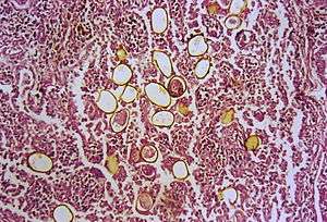
The acute phase (invasion and migration) may be marked by diarrhea, abdominal pain, fever, cough, urticaria, hepatosplenomegaly, pulmonary abnormalities, and eosinophilia. During the chronic phase, pulmonary manifestations include cough, expectoration of discolored sputum containing clumps of eggs,[3] hemoptysis, and chest radiographic abnormalities. Extrapulmonary locations of the adult worms result in more severe manifestations, especially when the brain is involved."[6] "Diagnosis is based on microscopic demonstration of eggs in stool or sputum, but these are not present until 2 to 3 months after infection. (Eggs are also occasionally encountered in effusion fluid or biopsy material.) Concentration techniques may be necessary in patients with light infections. Biopsy may allow diagnostic confirmation and species identification when an adult or developing fluke is recovered.[6]
Paragonimiasis can commonly be misdiagnosed as tuberculosis.[7]
Treatment
The drug of choice to treat paragonimiasis is praziquantel, although bithionol may also be used.[6]
See also
- Drunken shrimp
References
- Haswell-Elkins MR, Elkins DB (1998). "Lung and liver flukes". In Collier L, Balows A, Sussman M (eds.). Topley and Wilson's Microbiology and Microbial Infections. 5 (9th ed.). New York: Oxford University Press. pp. 507–520. ISBN 978-0340663202.
- "Paragonimiasis: Background, Pathophysiology, Epidemiology". 2016-06-20. Cite journal requires
|journal=(help) - "Paragonimiasis". Center for Global Health, U.S. Centers for Disease Control and Prevention (CDC). 2010-10-13. Retrieved 2012-09-06.
- "CDC - DPDx Homepage". 2019-02-04.
- "Foodborne trematode infections". World Health Organization. 2016. Retrieved November 11, 2016.
- "Paragonimiasis, Clinical Features". CDC. 2010-10-13. Retrieved 2012-09-06.
- Lane MA, Barsanti MC, Santos CA, Yeung M, Lubner SJ, Weil GJ (2009). "Human Paragonimiasis in North America following Ingestion of Raw Crayfish" (PDF). Clinical Infectious Diseases. 49 (6): e55–e61. doi:10.1086/605534. PMID 19681705.
External links
| Classification | |
|---|---|
| External resources |