Heterophyes heterophyes
| Heterophyes heterophyes | |
|---|---|
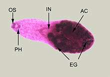 | |
| Adult specimen stained with carmine | |
| Scientific classification | |
| Kingdom: | Animalia |
| Phylum: | Platyhelminthes |
| Class: | Rhabditophora |
| Order: | Plagiorchiida |
| Family: | Heterophyidae |
| Genus: | Heterophyes |
| Species: | H. heterophyes |
| Binomial name | |
| Heterophyes heterophyes (Siebold, 1853) | |
| Synonyms | |
| |
Heterophyes heterophyes is a human parasite.
Distribution
This species occurs in Egypt, Sudan, Israel, Brazil, Spain, Turkey, Iran, India, and Russia.[1] Common in North Africa, Asia Minor, Korea, China, Japan, Taiwan, and the Philippines. In Egypt, after looking at twelve populations of Cerithideopsilla conica, which is mud snail and carrier of Heterophyes heterophyes, it was found that the most infected snails were found in the Nile Delta compared to the Red sea coast and inland waters of the Delta.[2] The transmission of Heterophyes heterophyes from, Pirenella conica, a snail, to fish which live in Northern Africa in lagoons that are connected to the sea. For transmission to occur from snails to fish the snails had to be larger than 5 mm.[3]
Morphology
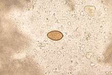
Minute teardrop-shaped flukes found in the small intestines of fish-eating birds and mammals. The eggs are hard to tell apart from other related species so there is no accurate estimate of human infection. The adult flukes range from 1.1 mm to 1.7 mm long and about 0.35 mm at their greatest width. The body of the fluke is covered in scales mostly concentrated at the anterior end. Also at the anterior end is an oral sucker. Located in the medioanterior of the body is the acetabulum. At the posterior end of the fluke are two oval testes. The vas deferens leading from the testes expands to form a seminal vesicle and then narrows again to form an ejaculatory duct. The fluke also has female reproductive organs. Located medioposterior is the fluke's one ovary and leading away from the ovary is the vitellaria. The uterus is a long tube like structure that also leads away from the ovary and joins up with the ejaculatory duct to form the genital duct which leads to a genital sinus. The sinus leads to the genital pore which is lined with 60-90 toothed spines. When a H. heterophyes was taken out of a man and then looked at the ultrastructures on the tegument it was described to have spines that looked like a round comb.[4] The genital pore is where the fluke releases its eggs. H. heterophyes will also have morphological differences based on the different fishes that it inhabited.[5]
Life cycle
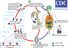
The adult flukes live burrowed between the villi of the host's small intestine. It only takes around 4 to 6 hours for H. heterophyes to get to the small intestines in the definitive host and even faster in hosts that it does not prefer.[6] The eggs that are laid contain a miracidium but do not hatch until they are ingested by a snail (Cerithideopsilla conica[1] in Egypt or Cerithidia cingula in Japan). Inside the snails gut, the miracidium becomes a sporocyst which then begin to produce rediae. The rediae produce cercariae which then exit the snail, swim toward the surface of the water, and slowly fall back down. On their way down, they contact a fish and penetrate into the epithelium of the fish. Here, the cercariae encyst in the muscle tissue. The second intermediate host include freshwater fish: Mugil cephalus, Tilapia nilotica, Aphanius fasciatus, and Acanthogobius sp.[1] The definitive host, such as humans or birds, eats the undercooked or raw meat of a fish and ingest the parasite. Natural definitive hosts are cats, dogs, foxes, wolves, pelicans, and humans.[1]
Epidemiology
People at high risk for infection are those who live by bay waters including fishermen. Infection is acquired by eating raw fish, a common food in areas of heavy endemicity. In endemic areas, people who live near lake shores or river banks usually have a higher rate and intensity of infection than those living a distance from such areas. It is possible that inhabitants of these areas eat more low-salted or improperly cooked fish and that their fish are obtained from polluted water. It is common practice for people to defecate on the lake shores and river banks or from their boats while fishing. In rural areas of Egypt there was a higher chance of being infected with H. heterophyes due to being a lower socioeconomic area and not having easy access to medical services.[7]
Pathology
Each worm causes a mild inflammatory reaction at its site of contact with the intestine. In heavy infections which are common cause damage to the mucosa and produce intestinal pain and mucosa diarrhea. Sometimes eggs can enter the blood and lymph vascular systems through mucosa go into the ectopic sites in the body. The heart can be affected with tissue reaction in the valves and myocardium that cause heart failure. Eggs can also get into the brain or spinal cord and cause neurological disorders and sometimes fatalities. Antigen and immune complex deposits left by H. heterophyes in the brain and kidneys of mice prove that there are changes in these tissues of the infected.[8]
Diagnosis
Diagnosis done by stool examination is difficult when adult worms are not present because the eggs are hard to distinguish from C.sinensis.
Treatment
Praziquantel, a quinolone derivative. The effect of praziquantel on H. heterophyes causes deep lesions on their teguments, and when exposed to praziquantel over a longer period of time leads to even deeper lesions.[9]
References
- Chai J. Y., Darwin Murrell K. & Lymbery A. J. (2005). "Fish-borne parasitic zoonoses: Status and issues". International Journal for Parasitology 35(11-12): 1233-1254. doi:10.1016/j.ijpara.2005.07.013.
- Taraschewski, H. (1985). Investigations on the prevalence of Heterophyes species in twelve populations of the first intermediate host in Egypt and Sudan. The Journal of Tropical Medicine and Hygiene, 88(4), 265-271.
- Taraschewski, H., & Paperna, I. (1981). Distribution of the snail Pirenella conica in Sinai and Israel and its infection by Heterophyidae and other trematodes. Marine Ecology, Progress Series, 5(2), 193-205.
- Uga, S., Morimoto, M., Saito, T., & Rai, S. K. (1999). Surface ultrastructure of Heterophyes heterophyes (Trematoda: Heterophyidae) collected from a man. JOURNAL-HELMINTHOLOGICAL SOCIETY WASHINGTON, 65, 119-121.
- El-Shazly, A. M., Soltan, D. M., El-sheikha, H. M., Morsy, G. H., & Morsy, T. A. (2007). Host-induced phenotypic differences in Egyptian Heterophyes heterophyes (Digenea: Heterophyidae). Journal of the Egyptian Society of Parasitology, 37(3), 815-824.
- Taraschewski, H. (1987). Experiments on habitat selection of Heterophyes species in different definitive hosts. Journal of Helminthology, 61, pp 33-42. doi:10.1017/S0022149X00009688.
- El-Shazly, A. M., El-Nahas, H. A., Soliman, M., Sultan, D. M., Abedl, T. A., & Morsy, T. A. (2006). The reflection of control programs of parasitic diseases upon gastrointestinal helminthiasis in Dakahlia Governorate, Egypt. Journal of the Egyptian Society of Parasitology, 36(2), 467-480.
- Daoud, A. (2012). Allergies in Other Diseases: 254 Experimental Heterophyiasis: Histopathological & Immunological Study. The World Allergy Organization Journal, 5(Suppl 2), S100.
- Taraschewski, H., Mehlhorn, H., Bunnag, D., Andrews, P., & Thomas, H. (1986). Effects of praziquantel on human intestinal flukes (Fasciolopsis buski and Heterophyes heterophyes). Zentralblatt für Bakteriologie, Mikrobiologie und Hygiene. Series A: Medical Microbiology, Infectious Diseases, Virology, Parasitology, 262(4), 542-550.
- Roberts, Larry, and John Janovy. Gerald D. Schmidt & Larry S. Roberts' Foundations of Parasitology. 8th ed. New York: McGraw-Hill, 2009. 291-92. Print.
| Wikimedia Commons has media related to Heterophyes heterophyes. |