Owl's eye appearance
Owl's eye appearance is a pattern in medical radiology and histopathology, and may refer to:
- Cells with perinuclear vacuolization around centrally located pyknotic nuclei, such as typically seen in flat warts.[1]
- Owl's eye appearance of inclusion bodies, which is highly specific for cytomegalovirus infection.[2]
- Owl's eye appearance of entire nucleus - a finding in Reed–Sternberg cells in individuals with Hodgkin's lymphoma.
- Owl's eye appearance of the Lentiform nucleus of the basal ganglia on head CT scan images in individuals with cerebral hypoxia[3]
.jpg)
The eye of an owl.
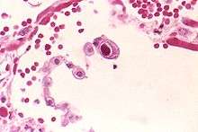
Cytomegalovirus infection of a lung pneumocyte, showing owl's eye appearance of a large cell at center.
References
- Ahmad M. Al Aboud; Pramod K. Nigam. "Wart (Plantar, Verruca Vulgaris, Verrucae)". StatPearls at National Center for Biotechnology Information. Last Update: May 13, 2019.
- Mattes FM, McLaughlin JE, Emery VC, Clark DA, Griffiths PD (August 2000). "Histopathological detection of owl's eye inclusions is still specific for cytomegalovirus in the era of human herpesviruses 6 and 7". J. Clin. Pathol. 53 (8): 612–4. doi:10.1136/jcp.53.8.612. PMC 1762915. PMID 11002765.
- http://radiographics.rsna.org/content/28/2/417.full
This article is issued from
Wikipedia.
The text is licensed under Creative
Commons - Attribution - Sharealike.
Additional terms may apply for the media files.