Ligament of head of femur
In human anatomy, the ligament of the head of the femur (round ligament of the femur, ligamentum teres femoris, or the foveal ligament) is a ligament located in the hip. It is triangular in shape and somewhat flattened. The ligament is implanted by its apex into the antero-superior part of the fovea capitis femoris and its base is attached by two bands, one into either side of the acetabular notch, and between these bony attachments it blends with the transverse ligament.[1]
| Ligament of head of femur | |
|---|---|
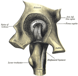 Left hip-joint, opened by removing the floor of the acetabulum from within the pelvis (Ligament of head of femur labeled as ligt. teres at cente.) | |
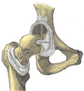 Hip-joint, front view. The capsular ligament has been largely removed (ligament visible at center labeled as ligam teres) | |
| Details | |
| From | Femur head |
| To | Acetabular notch |
| Identifiers | |
| Latin | Ligamentum capitis femoris, ligamentum teres femoris |
| MeSH | D000069593 |
| TA | A03.6.07.010 |
| FMA | 43235 |
| Anatomical terminology | |
It is ensheathed by the synovial membrane, and varies greatly in strength in different subjects; occasionally only the synovial fold exists, and in rare cases even this is absent.[1]
The ligament of the head of the femur contains within it the acetabular branch of the obturator artery.
Function
The ligament is made tense when the thigh is semiflexed and the limb then abducted or rotated outward; it is, on the other hand, relaxed when the limb is adducted.[1]
Research suggests it contributes little influence as a ligament past childhood,[2] although it may still be important in transmitting arterial supply to the femoral head.
In humans, it has been suggested that it is not the ligamentum teres but the hip capsule (specifically the iliofemoral, ischiofemoral and pubofemoral ligaments) that provides the primary resistance to dislocation in the extended hip. However, recent research has suggested the ligamentum teres of the femur may have a number of functions, including a significant biomechanical role on the basis of cadaveric studies where increases of range of motion were seen after sectioning of the ligament.[3]
Other animals
It has been suggested that some animals, such as the orangutan and Indian elephant, lack a ligamentum teres.[4][5] However, the presence of a ligamentum teres, albeit with a morphology different from the human version, has been found upon dissection in both these animals. In the orangutan, it is believed to play a significant role in preventing dislocation of the femoral head within extreme ranges of motion. In the Indian elephant, it is the primary support of the hip joint when the hind limbs are abducted.[6]
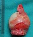 A human femur head with some synovium attached at the bottom and the ligament of the head of the femur attached at the top. A blue suture wire is drawn through the ligament. Ruler in centimetres at left side.
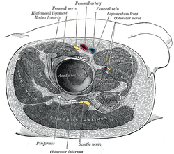 Structures surrounding right hip-joint
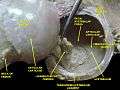 Hip joint. Lateral wiev. Ligament of head of femur
References
This article incorporates text in the public domain from page 336 of the 20th edition of Gray's Anatomy (1918)
- Gray's Anatomy (1918), see infobox
- Tan CK, Wong WC (August 1990). "Absence of the ligament of head of femur in the human hip joint". Singapore Medical Journal. 31 (4): 360–3. PMID 2124003.
- O'Donnell JM, Pritchard M, Salas AP, Singh PJ (July 2014). "The ligamentum teres-its increasing importance". Journal of Hip Preservation Surgery. 1 (1): 3–11. doi:10.1093/jhps/hnu003. PMC 4765261. PMID 27011796.
- Femur article, Encyclopædia Britannica.
- Ishida, Hidemi (2006). "Current Thoughts on Terrestrialization in African Apes and the Origin of Human Bipedalism". In Ishida, Hidemi; Tuttle, Russell; Pickford, Martin; Ogihara, Naomichi; Nakatsukasa, Masato (eds.). Human Origins and Environmental Backgrounds. Developments in Primatology: Progress and Prospects. pp. 259–66. doi:10.1007/0-387-29798-7_20. ISBN 9780387296388.
- Crelin ES. "Ligament of the head of the femur in the orangutan and Indian elephant". Yale J Biol Med. 61: 383–8. PMC 2590443. PMID 3201784.
| Authority control |
|---|