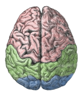
Lateralization of brain function
The lateralization of brain function is the tendency for some neural functions or cognitive processes to be specialized to one side of the brain or the other. The medial longitudinal fissure separates the human brain into two distinct cerebral hemispheres, connected by the corpus callosum. Although the macrostructure of the two hemispheres appears to be almost identical, different composition of neuronal networks allows for specialized function that is different in each hemisphere. Lateralization of brain structures is based on general trends expressed in healthy patients; however, there are numerous counterexamples to each generalization. Each human's brain develops differently leading to unique lateralization in individuals. This is different from specialization as lateralization refers only to the function of one structure divided between two hemispheres. Specialization is much easier to observe as a trend since it has a stronger anthropological history.[1] The best example of an established lateralization is that of Broca's and Wernicke's areas where both are often found exclusively on the left hemisphere. These areas frequently correspond to handedness, however, meaning that the localization of these areas is regularly found on the hemisphere corresponding to the dominant hand (anatomically on the opposite side). Function lateralization, such as semantics, intonation, accentuation, and prosody, has since been called into question and largely been found to have a neuronal basis in both hemispheres.[2] Another example is that each hemisphere in the brain tends to represent one side of the body. In the cerebellum this is the same bodyside, but in the forebrain this is predominantly the contralateral side.
Lateralized functions
Language
Language functions such as grammar, vocabulary and literal meaning are typically lateralized to the left hemisphere, especially in right-handed individuals.[3] While language production is left-lateralized in up to 90% of right-handers, it is more bilateral, or even right-lateralized, in approximately 50% of left-handers.[4]
Broca's area and Wernicke's area areas associated with the production of speech and comprehension of speech, respectively, are located in the left cerebral hemisphere for about 95% of right-handers, but about 70% of left-handers.[5]:69
Sensory processing
The processing of basic sensory information is lateralized by being divided into left and right sides of the body or the space around the body.
In vision, about half the neurons of the optic nerve from each eye cross to project to the opposite hemisphere and about half do not cross to project to the hemisphere on the same side.[6] This means that the left side of the visual field is processed largely by the visual cortex of the right hemisphere and vice versa for the right side of the visual field.
In audition, about 90% of the neurons of the auditory nerve from one ear cross to project to the auditory cortex of the opposite hemisphere.
In the sense of touch, most of the neurons from the skin cross to project to the somatosensory cortex of the opposite hemisphere.
Because of this functional division of the left and right sides of the body and of the space that surrounds it, the processing of information in the sensory cortices is essentially identical. That is, the processing of visual and auditory stimuli, spatial manipulation, facial perception, and artistic ability are represented bilaterally.[4] Numerical estimation, comparison and online calculation depend on bilateral parietal regions[7][8] while exact calculation and fact retrieval are associated with left parietal regions, perhaps due to their ties to linguistic processing.[7][8]
Value systems
Rather than just being a series of places where different brain modules occur, there are running similarities in the kind of function seen in each side, for instance how right-side impairment of drawing ability making patients draw the parts of the subject matter with wholly incoherent relationships, or where the kind of left-side damage seen in language impairment not damaging the patient's ability to catch the significance of intonation in speech.[9] This has led Iain McGilchrist to say that the two hemispheres as having different value systems, where the left hemisphere tends to reduce complex matters such as ethics to rules and measures, where the right hemisphere is disposed to the holistic and metaphorical.[10]
Clinical significance
Depression is linked with a hyperactive right hemisphere, with evidence of selective involvement in "processing negative emotions, pessimistic thoughts and unconstructive thinking styles", as well as vigilance, arousal and self-reflection, and a relatively hypoactive left hemisphere, "specifically involved in processing pleasurable experiences" and "relatively more involved in decision-making processes".[11] Additionally, "left hemisphere lesions result in an omissive response bias or error pattern whereas right hemisphere lesions result in a commissive response bias or error pattern."[12] The delusional misidentification syndromes, reduplicative paramnesia and Capgras delusion are also often the result of right hemisphere lesions.[13]
Hemisphere damage
Damage to either the right or left hemisphere, and its resulting deficits provide insight into the function of the damaged area. Left hemisphere damage has many effects on language production and perception. Damage or lesions to the right hemisphere can result in a lack of emotional prosody or intonation when speaking. Right hemisphere damage also has grave effects on understanding discourse. People with damage to the right hemisphere have a reduced ability to generate inferences, comprehend and produce main concepts, and a reduced ability to manage alternative meanings. Furthermore, people with right hemisphere damage often exhibit discourse that is abrupt and perfunctory or verbose and excessive. They can also have pragmatic deficits in situations of turn taking, topic maintenance and shared knowledge.
Lateral brain damage can also affect visual perceptual spatial resolution. People with left hemisphere damage may have impaired perception of high resolution, or detailed, aspects of an image. People with right hemisphere damage may have impaired perception of low resolution, or big picture, aspects of an image.
Plasticity
If a specific region of the brain, or even an entire hemisphere, is injured or destroyed, its functions can sometimes be assumed by a neighboring region in the same hemisphere or the corresponding region in the other hemisphere, depending upon the area damaged and the patient's age.[14] When injury interferes with pathways from one area to another, alternative (indirect) connections may develop to communicate information with detached areas, despite the inefficiencies.
Broca's aphasia
Broca's aphasia is a specific type of expressive aphasia and is so named due to the aphasia that results from damage or lesions to the Broca's area of the brain, that exists most commonly in the left inferior frontal hemisphere. Thus, the aphasia that develops from the lack of functioning of the Broca's area is an expressive and non-fluent aphasia. It is called 'non-fluent' due the issues that arise because Broca's area is critical for language pronunciation and production. The area controls some motor aspects of speech production and articulation of thoughts to words and as such lesions to the area result in the specific non-fluent aphasia.[15]
Wernicke's aphasia
Wernicke's aphasia is the result of damage to the area of the brain that is commonly in the left hemisphere above the sylvian fissure. Damage to this area causes primarily a deficit in language comprehension. While the ability to speak fluently with normal melodic intonation is spared, the language produced by a person with Wernicke's aphasia is riddled with semantic errors, and may sound nonsensical to the listener. Wernicke's aphasia is characterized by phonemic paraphasias, neologism or jargon. Another characteristic of a person with Wernicke's aphasia is that they are unconcerned by the mistakes that they are making.
Society and culture
Misapplication
Terence Hines states that the research on brain lateralization is valid as a research program, though commercial promoters have applied it to promote subjects and products far outside the implications of the research.[16] For example, the implications of the research have no bearing on psychological interventions such as EMDR and neurolinguistic programming,[17] brain-training equipment, or management training.[18]
Pop psychology

Some popularizations oversimplify the science about lateralization, by presenting the functional differences between hemispheres as being more absolute than is actually the case.[19]:107[20]
Sex differences
In the 19th century and to a lesser extent the 20th, it was thought that each side of the brain was associated with a specific gender: the left corresponding with masculinity and the right with femininity and each half could function independently.[21] The right side of the brain was seen as the inferior and thought to be prominent in women, savages, children, criminals, and the insane. A prime example of this in fictional literature can be seen in Robert Louis Stevenson's Strange Case of Dr. Jekyll and Mr. Hyde.[22]
Evolutionary advantage
The widespread lateralization of many vertebrate animals indicates an evolutionary advantage associated with the specialization of each hemisphere.[1]
History
Broca
One of the first indications of brain function lateralization resulted from the research of French physician Pierre Paul Broca, in 1861. His research involved the male patient nicknamed "Tan", who suffered a speech deficit (aphasia); "tan" was one of the few words he could articulate, hence his nickname. In Tan's autopsy, Broca determined he had a syphilitic lesion in the left cerebral hemisphere. This left frontal lobe brain area (Broca's area) is an important speech production region. The motor aspects of speech production deficits caused by damage to Broca's area are known as expressive aphasia. In clinical assessment of this aphasia, it is noted that the patient cannot clearly articulate the language being employed.
Wernicke
German physician Karl Wernicke continued in the vein of Broca's research by studying language deficits unlike expressive aphasia. Wernicke noted that not every deficit was in speech production; some were linguistic. He found that damage to the left posterior, superior temporal gyrus (Wernicke's area) caused language comprehension deficits rather than speech production deficits, a syndrome known as receptive aphasia.
Imaging
These seminal works on hemispheric specialization were done on patients or postmortem brains, raising questions about the potential impact of pathology on the research findings. New methods permit the in vivo comparison of the hemispheres in healthy subjects. Particularly, magnetic resonance imaging (MRI) and positron emission tomography (PET) are important because of their high spatial resolution and ability to image subcortical brain structures.
Movement and sensation
In the 1940s, neurosurgeon Wilder Penfield and his neurologist colleague Herbert Jasper developed a technique of brain mapping to help reduce side effects caused by surgery to treat epilepsy. They stimulated motor and somatosensory cortices of the brain with small electrical currents to activate discrete brain regions. They found that stimulation of one hemisphere's motor cortex produces muscle contraction on the opposite side of the body. Furthermore, the functional map of the motor and sensory cortices is fairly consistent from person to person; Penfield and Jasper's famous pictures of the motor and sensory homunculi were the result.
Split-brain patients
Research by Michael Gazzaniga and Roger Wolcott Sperry in the 1960s on split-brain patients led to an even greater understanding of functional laterality. Split-brain patients are patients who have undergone corpus callosotomy (usually as a treatment for severe epilepsy), a severing of a large part of the corpus callosum. The corpus callosum connects the two hemispheres of the brain and allows them to communicate. When these connections are cut, the two halves of the brain have a reduced capacity to communicate with each other. This led to many interesting behavioral phenomena that allowed Gazzaniga and Sperry to study the contributions of each hemisphere to various cognitive and perceptual processes. One of their main findings was that the right hemisphere was capable of rudimentary language processing, but often has no lexical or grammatical abilities.[23] Eran Zaidel also studied such patients and found some evidence for the right hemisphere having at least some syntactic ability.
Language is primarily localized in the left hemisphere. One of the experiments carried out by Gazzaniga involved a split-brain male patient sitting in front of a computer screen while having words and images presented on either side of the screen and the visual stimuli would go to either the right or left visual field, and thus the left or right brain, respectively. It was observed that if the patient was presented with an image to his left visual field (right brain), he would report not seeing anything. If he was able to feel around for certain objects, he could accurately pick out the correct object, despite not having the ability to verbalize what he saw. This led to confirmation that the left brain is localized for language whereas the right brain does not have this capability, and when the corpus callosum is cut, the two hemispheres cannot communicate in order for situation-pertinent speech to be produced.
Additional images
 Ventricles of brain and basal ganglia. Superior view. Horizontal section. Deep dissection
 Ventricles of brain and basal ganglia. Superior view. Horizontal section. Deep dissection
See also
- Alien hand syndrome
- Ambidexterity
- Bicameralism
- Brain asymmetry
- Chirality
- Contralateral brain
- Cross-dominance
- Divided consciousness
- Dual consciousness
- Emotional lateralization
- Handedness
- Hemispherectomy
- Laterality
- Left brain interpreter
- Parallel computing
- Psychoneuroimmunology
- Right hemisphere brain damage
- Wada test
- Yakovlevian torque
References
- Halpern ME, Güntürkün O, Hopkins WD, Rogers LJ (2005). "Lateralization of the Vertebrate Brain: Taking the Side of Model Systems". The Journal of Neuroscience. 25 (45): 10351–10357. doi:10.1523/JNEUROSCI.3439-05.2005. PMC 2654579. PMID 16280571.
- Riès, SK; Dronkers, NF; Knight, RT (April 2016). "Choosing words: left hemisphere, right hemisphere, or both? Perspective on the lateralization of word retrieval". Annals of the New York Academy of Sciences. 1369 (1): 111–31. doi:10.1111/nyas.12993. PMC 4874870. PMID 26766393.
- Taylor, I.; Taylor, M. M. (1990). Psycholinguistics: Learning and using Language. Pearson. ISBN 978-0-13-733817-7. p. 367
- Beaumont, J.G. (2008). Introduction to Neuropsychology, Second Edition. The Guilford Press. ISBN 978-1-59385-068-5. Chapter 7
- Griggs, Richard A. (2012). Psychology : a concise introduction (3rd ed.). New York, NY: Worth Publishers. ISBN 978-1429261555.
- https://theodora.com/anatomy/the_optic_nerve.html
- Dehaene S, Spelke E, Pinel P, Stanescu R, Tsivkin S (May 1999). "Sources of mathematical thinking: behavioral and brain-imaging evidence" (PDF). Science. 284 (5416): 970–4. doi:10.1126/science.284.5416.970. PMID 10320379. Archived (PDF) from the original on 19 July 2011.
- Dehaene S, Piazza M, Pinel P, Cohen L (2003). "Three parietal circuits for number processing" (PDF). Cognitive Neuropsychology. 20 (3–6): 487–506. CiteSeerX 10.1.1.4.8178. doi:10.1080/02643290244000239. PMID 20957581. Archived (PDF) from the original on 19 July 2011.
- McGilchrist (2009) provides an extensive survey of the relevant literature in chapter two.
- Iain McGilchrist & Shankar Vedantam (4 February 2019). "One Head, Two Brains: How The Brain's Hemispheres Shape The World We See" (Audio podcast with transcript). NPR Hidden Brain.
- Hecht D (October 2010). "Depression and the hyperactive right-hemisphere". Neurosci. Res. 68 (2): 77–87. doi:10.1016/j.neures.2010.06.013. PMID 20603163.
- Braun CM, Delisle J, Guimond A, Daigneault R (March 2009). "Post unilateral lesion response biases modulate memory: crossed double dissociation of hemispheric specialisations". Laterality. 14 (2): 122–64. doi:10.1080/13576500802328613. PMID 18991140.
- Devinsky O (January 2009). "Delusional misidentifications and duplications: right brain lesions, left brain delusions". Neurology. 72 (1): 80–7. doi:10.1212/01.wnl.0000338625.47892.74. PMID 19122035.
- Pulsifer MB, Brandt J, Salorio CF, Vining EP, Carson BS, Freeman JM (2004). "The cognitive outcome of hemispherectomy in 71 children". Epilepsia. 45 (3): 243–254. doi:10.1111/j.0013-9580.2004.15303.x. PMID 15009226.
- Biopsychology (8th edition), by John J.P. Pinel Pearson 2011
- Hines, Terence (1987). "Left Brain/Right Brain Mythology and Implications for Management and Training". The Academy of Management Review. 12 (4): 600–606. doi:10.2307/258066. JSTOR 258066.
- Drenth JD (2003). "Growing anti-intellectualism in Europe; a menace to science". Studia Psychologica. 45 (1): 5–13., available in ALLEA Annual Report 2003 Archived 16 June 2011 at the Wayback Machine, pp. 61–72
- Della Sala, Sergio (1999). Mind Myths: Exploring Popular Assumptions about the Mind and Brain. New York: Wiley. ISBN 978-0-471-98303-3. Archived from the original on 9 May 2018.
- Westen, Drew; Burton, Lorelle; Kowalski, Kowalski (2006). Psychology : Australian and New Zealand edition. Milton, Qld.: John Wiley & Sons. ISBN 9780470805527.
- Toga AW, Thompson PM (2003). "Mapping brain asymmetry". Nature Reviews Neuroscience. 4 (1): 37–48. doi:10.1038/nrn1009. PMID 12511860.
- Harrington, Anne (1 January 1989). Medicine, Mind, and the Double Brain: A Study in Nineteenth-Century Thought. Princeton University Press. pp. 87–90. ISBN 978-0691024226.
- Stiles, Anne (2006). "Robert Louis Stevenson's "Jekyll and Hyde" and the Double Brain". SEL: Studies in English Literature 1500–1900. 46 (4): 879–900. JSTOR 4127513.
- Kandel E, Schwartz J, Jessel T. Principles of Neural Science. 4th ed. p1182. New York: McGraw–Hill; 2000. ISBN 0-8385-7701-6
Bibliography
- McGilchrist, Iain (9 October 2009). The Master and His Emissary: The Divided Brain and the Making of the Western World (Hardcover ed.). USA: Yale University Press. ISBN 978-0-300-14878-7.
Further resources
- Josse G, Tzourio-Mazoyer N (2003). "Review: Hemispheric specialization for language". Brain Research Reviews. 44 (1): 1–12. doi:10.1016/j.brainresrev.2003.10.001. PMID 14739000.
- Cutting, John (2012). A Critique of Psychopathology. Parodos Verlag. ISBN 978-3-938880-51-7.
- Ornstein, Robert (1998). The Right Mind: Making Sense of the Hemispheres. Harcourt Brace International. ISBN 978-0-15-600627-9.
- Jill Bolte Taylor (2008). My Stroke of Insight. Viking: USA. 0670020745.