Neuroscience of sex differences
The neuroscience of sex differences is the study of characteristics that separate the male and female brain. Psychological sex differences are thought by some to reflect the interaction of genes, hormones and social learning on brain development throughout the lifespan.
| This article is one of a series on: |
| Sex differences in humans |
|---|
| Physiology |
| Medicine |
|
| Neuroscience |
|
| Sociology |
|
Some evidence from brain morphology and function studies indicates that male and female brains cannot always be assumed to be identical from either a structural or functional perspective, and some brain structures are sexually dimorphic.[1][2]
History
The ideas of differences between the male and female brain have circulated since the time of ancient Greek philosophers around 850 B.C. In 1854, Emil Huschke discovered a size difference in the frontal lobe, where male frontal lobes are 1% larger than females.[3] As the 19th century progressed, scientists began researching sexual dimorphisms in the brain significantly more.[4] Until recent decades, scientists knew of several structural sexual dimorphisms of the brain, but they did not think that sex had any impact on how the human brain performs daily tasks. Through molecular, animal, and neuroimaging studies, a great deal of information regarding the differences between male and female brains and how much they differ in regards to both structure and function has been uncovered.[5]
Evolutionary explanations
Sexual selection
Females show enhanced information recall compared to males. This may be due to the fact that females have a more intricate evaluation of risk-scenario contemplation, based on a prefrontal cortical control of the amygdala. For example, the ability to recall information better than males most likely originated from sexual selective pressures on females during competition with other females in mate selection. Recognition of social cues was an advantageous characteristic because it ultimately maximized offspring and was therefore selected for during evolution.[1]
Oxytocin is a hormone that induces contraction of the uterus and lactation in mammals and is also a characteristic hormone of nursing mothers. Studies have found that oxytocin improves spatial memory. Through activation of the MAP kinase pathway, oxytocin plays a role in the enhancement of long-term synaptic plasticity, which is a change in strength between two neurons over a synapse that lasts for minutes or longer, and long-term memory. This hormone may have helped mothers remember the location of distant food sources so they could better nurture their offspring.[1]
Male and female brain anatomy
Males and females differ in some aspects of their brains, notably the overall difference in size with men having larger brains on average (between 8% and 13% larger),[2] but there are areas of the brain which appear not to be sexually differentiated. Additionally, there are differences in activation patterns which suggest anatomical or developmental differences, but the source of these differences is often unclear.
Lateralization
Lateralization may differ between the sexes with men often being said to have a more lateralized brain. This is often attributed to differences in "left" and "right" brained abilities. One factor which contributes support to the idea that there is a sex difference in brain lateralization is that men are more likely to be left handed. However, it is unclear whether this is due to a difference in lateralization.[6]
Meta-analysis of grey matter in the brain found sexually dimorphic areas of the brain in both volume and density. When synthesized, these differences show volume increases for males tend to be on the left side of systems, while females generally see greater volume in the right hemisphere.[2] However, based on a number of studies using different measurement techniques, there is no significant difference between male and female brain lateralization.[6]
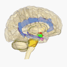
Amygdala
There are behavioral differences between males and females that may suggest a difference in amygdala size or function. Based on a review of amygdala volume studies, there was a raw size difference with males having a 10% larger amygdala, however, because male brains are larger, this finding is misleading. After normalizing for brain size, there was no significant difference in size of the amygdala across sex.[7]
In terms of activation, there is no difference in amygdala activation across sex. Differences in behavioral tests may be due to potential anatomical and physiological differences in the amygdala across sexes rather than activation differences.[8]
Emotional expression, understanding, and behavior appears to vary between males and females, however, these differences have not been correlated to any difference in brain structure or size. A 2012 review concluded that males and females have differences in the processing of emotions. Males tend to have stronger reactions to threatening stimuli and that males react with more physical aggression, however no conclusion about the direct role of the amygdala was made.[9]
Hippocampus
Hippocampus atrophy is associated with a variety of psychiatric disorders which have higher prevalence in females. Additionally, there are differences in memory skills between males and females which may suggest a difference in the hippocampal volume (HCV). A meta-analysis of volume differences found a higher HCV in males without correcting for total brain size. However, after adjusting for individual differences and total brain volume, they found no sex difference despite the expectation that women may have larger hippocampus volume.[10]
Grey matter
The specific areas where differences were measured included males having more grey matter volume in the bilateral amygdala, hippocampus, and anterior parahippocampal gyri, among others, while females had more grey matter volume in the right frontal pole, inferior and middle frontal gyrus, anterior cingulate gyrus, and lateral occipital cortex, among others. In terms of density, there are also differences between the sexes. Males tend to have denser left amygdala, hippocampus, and areas of the right VI lobe of the cerebellum, among other areas, while females tend to have denser left frontal pole.[2] The significance of these differences lies both in the lateralization (males having more volume in left hemisphere and females having more volume in the right hemisphere) and the possible uses of these findings to explore differences in neurological and psychiatric conditions.
Transgender studies on brain anatomy
Early postmortem studies of transsexual neurological differentiation was focused on the hypothalamic and amygdala region of the brain. Using magnetic resonance imaging (MRI), some trans women were found to have female typical putamen that were larger in size than cisgender males.[11] Some trans women have also shown a female typical central part of the bed nucleus of the stria terminalis (BSTc) and interstitial nucleus of the anterior hypothalamus number 3 (INAH-3), looking at the number of neurons found within each.[12]
Brain networks
Both males and females have consistent active working memory networks composed of bilateral middle frontal gyri, left cingulate gyrus, right precuneus, left inferior and superior parietal lobes, right claustrum, and left middle temporal gyrus.[13] Although the same brain networks are used for working memory, specific regions are sex specific. Sex differences were evident in other networks as women also tend to have higher activity in the prefrontal and limbic regions, such as the anterior cingulate, bilateral amygdala, and right hippocampus, while men tend to have a distributed network spread out among the cerebellum, portions of the superior parietal lobe, the left insula, and bilateral thalamus.[13]
A 2017 review from the perspective of large-scale brain networks, hypothesized that women's higher susceptibility to stress-prone diseases like PTSD and major depressive disorder, in which the salience network is theorized to be overactive and to interfere with the executive control network, may be due in part, along with societal exposure to stressors and the coping strategies that are available to women, to underlying sex-based brain differences.[14]
Neurochemical differences
Hormones
Gonadal hormones, or sex hormones, include androgens (such as testosterone) and estrogens (such as estradiol) which are steroid hormones synthesized primarily in the testes and ovaries, respectively. Sex hormone production is regulated by the gonadotropic hormones luteinizing hormone (LH) and follicle-stimulating hormone (FSH), whose release from the anterior pituitary is stimulated by gonadotropin-releasing hormone (GnRH) from the hypothalamus.[15]
Steroid hormones have several effects on brain development as well as maintenance of homeostasis throughout adulthood. Estrogen receptors have been found in the hypothalamus, pituitary gland, hippocampus, and frontal cortex, indicating the estrogen plays a role in brain development. Gonadal hormone receptors have also been found in the basal fore-brain nuclei.[16]
Estrogen and the female brain
Estradiol influences cognitive function, specifically by enhancing learning and memory in a dose-sensitive manner. Too much estrogen can have negative effects by weakening performance of learned tasks as well as hindering performance of memory tasks; this can result in females exhibiting poorer performance of such tasks when compared to males.[17]
Ovariectomies, surgeries inducing menopause, or natural menopause cause fluctuating and decreased estrogen levels in women. This in turn can "attenuate the effects" of endogenous opioid peptides. Opioid peptides are known to play a role in emotion and motivation. β-endorphin (β-EP), an endogenous opioid peptide, content has been found to decrease (in varying amounts/brain region), post ovariectomy, in female rats within the hypothalamus, hippocampus, and pituitary gland. Such a change in β-EP levels could be the cause of mood swings, behavioral disturbances, and hot flashes in post menopausal women.[16]
Progesterone and the male and female brain
Progesterone is a steroid hormone synthesized in both male and female brains. It contains characteristics found in the chemical nucleus of both estrogen and androgen hormones.[18] As a female sex hormone, progesterone is more significant in females than in males. During the menstrual cycle, progesterone increases just after the ovulatory phase to inhibit luteinizing hormones, such as oxytocin absorption.[19] In men, increased progesterone has been linked to adolescents with suicidal ideation.[20]
Testosterone and the male brain
The gonadal hormone testosterone is an androgenic, or masculinizing, hormone that is synthesized in both the male testes and female ovaries,[21] at a rate of about 14000 μg/day and 600 μg/day, respectively.[15] Testosterone exerts organizational effects on the developing brain, many of which are mediated through estrogen receptors following its conversion to estrogen by the enzyme aromatase within the brain.[15]
Cognitive tasks
It was once thought that sex differences in cognitive task and problem solving did not occur until puberty. However, as of 2000, evidence suggested that cognitive and skill differences are present earlier in development. For example, researchers have found that three- and four-year-old boys were better at targeting and at mentally rotating figures within a clock face than girls of the same age were. Prepubescent girls, however, excelled at recalling lists of words. These sex differences in cognition correspond to patterns of ability rather than overall intelligence. Laboratory settings are used to systematically study the sexual dimorphism in problem solving task performed by adults.[22]
On average, males excel relative to females at certain spatial tasks. Specifically, males have an advantage in tests that require the mental rotation or manipulation of an object.[23] In a computer simulation of a maze task, males completed the task faster and with fewer errors than their female counterparts. Additionally, males have displayed higher accuracy in tests of targeted motor skills, such as guiding projectiles.[22] Males are also faster on reaction time and finger tapping tests.[24]
On average, females excel relative to males on tests that measure recollection. They have an advantage on processing speed involving letters, digits and rapid naming tasks.[24] Females tend to have better object location memory and verbal memory.[25] They also perform better at verbal learning.[26] Females have better performance at matching items and precision tasks, such as placing pegs into designated holes. In maze and path completion tasks, males learn the goal route in fewer trials than females, but females remember more of the landmarks presented. This shows that females use landmarks in everyday situations to orient themselves more than males. Females are better at remembering whether objects had switched places or not.[22]
See also
- Biology and sexual orientation
- Etiology of transsexualism
- Neuroplasticity
- Neuroscience and sexual orientation
- Neuroscience
- Puberty § Neurohormonal process
- Rett syndrome
- Sex differences in human psychology
- Sexual dimorphism
- Sexually dimorphic nucleus
- The NeuroGenderings Network
References
- Cahill L (June 2006). "Why sex matters for neuroscience". Nature Reviews. Neuroscience. 7 (6): 477–84. doi:10.1038/nrn1909. PMID 16688123.
- Ruigrok AN, Salimi-Khorshidi G, Lai MC, Baron-Cohen S, Lombardo MV, Tait RJ, Suckling J (February 2014). "A meta-analysis of sex differences in human brain structure". Neuroscience and Biobehavioral Reviews. 39: 34–50. doi:10.1016/j.neubiorev.2013.12.004. PMC 3969295. PMID 24374381.
- Swaab DF, Hofman MA (1984). Sexual differentiation of the human brain. A historical perspective. Progress in Brain Research. 61. pp. 361–74. doi:10.1016/S0079-6123(08)64447-7. ISBN 9780444805324. PMID 6396708.
- Hofman MA, Swaab DF (1991). "Sexual dimorphism of the human brain: myth and reality" (PDF). Experimental and Clinical Endocrinology. 98 (2): 161–70. doi:10.1055/s-0029-1211113. PMID 1778230.
- McCarthy MM (February 2016). "Multifaceted origins of sex differences in the brain". Philosophical Transactions of the Royal Society of London. Series B, Biological Sciences. 371 (1688): 20150106. doi:10.1098/rstb.2015.0106. PMC 4785894. PMID 26833829.
- Sommer IE, Aleman A, Somers M, Boks MP, Kahn RS (April 2008). "Sex differences in handedness, asymmetry of the planum temporale and functional language lateralization". Brain Research. 1206: 76–88. doi:10.1016/j.brainres.2008.01.003. PMID 18359009.
- Marwha D, Halari M, Eliot L (February 2017). "Meta-analysis reveals a lack of sexual dimorphism in human amygdala volume". NeuroImage. 147: 282–294. doi:10.1016/j.neuroimage.2016.12.021. PMID 27956206.
- Sergerie K, Chochol C, Armony JL (2008). "The role of the amygdala in emotional processing: a quantitative meta-analysis of functional neuroimaging studies". Neuroscience and Biobehavioral Reviews. 32 (4): 811–30. doi:10.1016/j.neubiorev.2007.12.002. PMID 18316124.
- Kret ME, De Gelder B (June 2012). "A review on sex differences in processing emotional signals" (PDF). Neuropsychologia. 50 (7): 1211–21. doi:10.1016/j.neuropsychologia.2011.12.022. PMID 22245006.
- Tan A, Ma W, Vira A, Marwha D, Eliot L (January 2016). "The human hippocampus is not sexually-dimorphic: Meta-analysis of structural MRI volumes". NeuroImage. 124 (Pt A): 350–366. doi:10.1016/j.neuroimage.2015.08.050. PMID 26334947.
- Saleem F, Rizvi SW (December 2017). "Transgender Associations and Possible Etiology: A Literature Review". Cureus. 9 (12): e1984. doi:10.7759/cureus.1984. PMC 5825045. PMID 29503778.
- Guillamon A, Junque C, Gómez-Gil E (October 2016). "A Review of the Status of Brain Structure Research in Transsexualism". Archives of Sexual Behavior. 45 (7): 1615–48. doi:10.1007/s10508-016-0768-5. PMC 4987404. PMID 27255307.
- Hill AC, Laird AR, Robinson JL (October 2014). "Gender differences in working memory networks: a BrainMap meta-analysis" (PDF). Biological Psychology. 102: 18–29. doi:10.1016/j.biopsycho.2014.06.008. PMC 4157091. PMID 25042764.
- Homberg JR, Kozicz T, Fernandez G (April 2017). "Large-scale network balances in the transition from adaptive to maladaptive stress responses". Current Opinion in Behavioral Sciences. 14: 27–32. doi:10.1016/j.cobeha.2016.11.003.
- Molina PE (2018). Endocrine physiology (5th ed.). New York: McGraw-Hill Education. ISBN 9781260019360. OCLC 1026417940.
- Genazzani AR, Pluchino N, Luisi S, Luisi M (2007). "Estrogen, cognition and female ageing". Human Reproduction Update. 13 (2): 175–87. doi:10.1093/humupd/dml042. PMID 17135285.
- Korol DL (November 2004). "Role of estrogen in balancing contributions from multiple memory systems". Neurobiology of Learning and Memory. 82 (3): 309–23. doi:10.1016/j.nlm.2004.07.006. PMID 15464412.
- Funk and Wagnalls (2018). Progesterone. World Almanac Education Group.
- Ulshöfer, Gotlind; Karafyllis, Nicole (2008). Sexualized Brains: Scientific Modeling of Emotional Intelligence from a Cultural Perspective. Cambridge, Massachusetts: The MIT Press. p. 213.
- Lester, David; Gunn, John F. III; Quinnett, Paul, eds. (2014). Suicide in Men: How Men Differ from Women in Expressing their Distress. Springfield, Illinois: Charles C Thomas. p. 61. ISBN 978-0-398-08794-4.
- Hadley ME, Levine JE (2007). Endocrinology. Levine, Jon E. (6th, New ed.). Upper Saddle River, NJ: Pearson Prentice Hall. ISBN 978-0131876064. OCLC 70929277.
- Kimura D (July 31, 2000). Sex and Cognition. A Bradford Book. p. 28. ISBN 978-0262611640.
- Miller DI, Halpern DF (January 2014). "The new science of cognitive sex differences". Trends in Cognitive Sciences. 18 (1): 37–45. doi:10.1016/j.tics.2013.10.011. PMID 24246136.
- Roivainen, Eka (2011). "Gender differences in processing speed: A review of recent research". Learning and Individual Differences. 21 (2): 145–149. doi:10.1016/j.lindif.2010.11.021.
- Li R (September 2014). "Why women see differently from the way men see? A review of sex differences in cognition and sports". Journal of Sport and Health Science. 3 (3): 155–162. doi:10.1016/j.jshs.2014.03.012. PMC 4266559. PMID 25520851.
- Wallentin M (March 2009). "Putative sex differences in verbal abilities and language cortex: a critical review". Brain and Language. 108 (3): 175–83. doi:10.1016/j.bandl.2008.07.001. PMID 18722007.
Further reading
- Rippon, Gina (28 Feb 2019). The gendered brain : the new neuroscience that shatters the myth of the female brain. Bodley Head. ISBN 978-1847924759.