Intermammary cleft
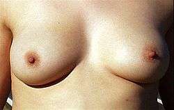
In female human anatomy, the intermammary cleft or intermammary sulcus or sulcus intermammarius of a woman is delineated by where the fatty portions of each breast sits in relationship to the sternum (or breastbone).[1] It divides the two mammary complexes that consist of two bodies of fatty pads, glandular tissues of mammary glands, connective tissues, and skin, as well as two duct systems (lactiferous duct of lymphatic vessel) and lobule alveoli emanating from two nipples.[2][3] Lymph vessels can ventrally extend as far as the intermammary sulcus.[4]
| Intermammary cleft | |
|---|---|
 Anatomical region of woman's cleavage | |
| Details | |
| Identifiers | |
| Latin | sulcus intermammarius |
| TA | A16.0.02.002 |
| FMA | 55264 |
| Anatomical terminology | |
Anatomy
.png)
The International Federation of Associations of Anatomists (IFAA) uses the terms "intermammary sulcus" or "intermammary cleft" when referring to the area between the breasts not including the breasts. In popular usage the area is commonly referred to as a woman's cleavage, especially when exposed by garments with low necklines.
For legal purposes it was noted by the United States federal courts that "anal cleft or cleavage" and "cleavage of the female breast" are so imprecise as to provide no guidance in defining them.[5] Medically, the "width" of a woman's cleavage is determined by the attachment points of her breast tissue to the periosteal tissue covering her sternum and is also defined somewhat by the medial attachments of the pectoralis major muscle when implants are in the sub-muscular position.[6]
Plastic surgeon Alan Matarasso said, "Ergonomically speaking, cleavage equals the position of the breasts on the chest wall."[7] The skin of the cleavage area is frailer than the skin of the face as it has fewer oil glands, and may show loss of elasticity sooner.[7]
Pathology
Poikiloderma of Civatte, a condition of dilated blood vessels and red to red-brown spots, is common to the upper part of the cleavage, especially for those who wear a sports bra or push-up bra for prolonged periods, and commonly affects fair skinned middle-aged to elderly women.[7][8]
It is characterized by hypopigmentation, hyperpigmentation, telangiectasias and superficial skin atrophy (occasional itching is reported), is another condition caused by long exposure to sunlight.[9][10]
Intermammary cleft can get attacked by plaque type psoriasis, which can in turn can cause erythematosus.[11]
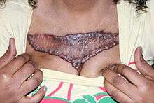
Hidradenitis suppurativa (HS) is a non-contagious chronic skin disease which affects intertriginous skin of apocrine sweat gland bearing areas like the inframammary fold and intermammary sulcus. It is characterized by clusters of abscesses, epidermoid cysts, sebaceous cysts, pilonidal cysts.[12][13]
Prurigo pigmentosa is a rare skin condition of unknown cause that affects depressed places on the chest and back like the intermammary area. It is characterized by the sudden onset of erythematous papules that leave a reticulated hyperpigmentation when they heal.[14][15]
Treatment
Poikiloderma and Dermatoheliosis are treated by desquamation (skin peeling).[7][9] There is no single effective treatment for HS. The recommended treatments include antibiotics, antiandrogens, corticosteroids, ciclosporins, and TNF inhibitors.[13]
References
- Dr. Ted Eisenberg and Joyce K. Eisenberg, ‘’ The Scoop on Breasts: A Plastic Surgeon Busts the Myths’’, Incompra Press, 2012, ISBN 978-0-9857249-3-1
- Heide Schatten and Gheorghe M. Constantinescu, Comparative Reproductive Biology, page 17, John Wiley & Sons, 2008, ISBN 978-0-470-39025-2
- Genaro Andres Contreras, The Use of Tylosin to Treat Intramammary Infections , page 22, ProQuest, 2008, ISBN 978-0-549-60762-5
- Charles Wesley Turner, The mammary gland (vol. 1), page 80, Lucas Bros., 1952
- West's federal supplement (First Series), p. 994, West Publishing Co, 1990
- Oliver, Rob (August 29, 2007). "I'll boost your boobs or go bust!". Retrieved 13 April 2013.
- Daphne Merkin, "The Great Divide", New York Times, August 28, 2005
- Rapini, Ronald P.; Bolognia, Jean L.; Jorizzo, Joseph L. (2007). Dermatology: 2-Volume Set. St. Louis: Mosby. ISBN 1-4160-2999-0.
- Murad Alam and Marisa Pongprutthipan, Body Rejuvenation, page 39, Springer Science & Business Media, 2010, ISBN 978-1-4419-1093-6
- American Osteopathic College of Dermatology "Dermatologic Disease Database", aocd.org, referenced July 22, 2011.
- Alan Menter and Benjamin Stoff, Psoriasis, page 25, CRC Press, 2010, ISBN 978-1-84076-590-8
- Alikhan, Ali; Lynch, Eisen (2009). "Hidradenitis suppurativa: a comprehensive review". J Am Acad Dermatol. 60 (4): 539–561. doi:10.1016/j.jaad.2008.11.911. PMID 19293006.
- Faye Lyons, Dermatology for the Advanced Practice Nurse, pages 118-121, Springer Publishing Company, 2014, ISBN 978-0-8261-3643-5
- James, William; Berger, Timothy; Elston, Dirk (2005). Andrews' Diseases of the Skin: Clinical Dermatology. (10th ed.). page 57, Saunders. ISBN 0-7216-2921-0.
- Q Ashton Acton, Issues in Dermatology and Cosmetic Medicine (2013 Edition), page 432, Scholarly Editions, 2013, ISBN 978-1-4901-0911-4