Internal iliac vein
The internal iliac vein (hypogastric vein) begins near the upper part of the greater sciatic foramen, passes upward behind and slightly medial to the internal iliac artery and, at the brim of the pelvis, joins with the external iliac vein to form the common iliac vein.
| Internal iliac vein | |
|---|---|
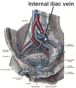 The veins of the right half of the male pelvis. | |
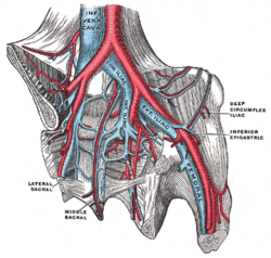 The iliac veins. (Int. iliac visible at center.) | |
| Details | |
| Drains to | Common iliac vein |
| Artery | Internal iliac artery |
| Identifiers | |
| Latin | Vena iliaca interna, vena hypogastrica |
| TA | A12.3.10.004 |
| FMA | 18884 |
| Anatomical terminology | |
Structure
Several veins unite above the greater sciatic foramen to form the internal iliac vein. It does not have the predictable branches of the internal iliac artery but its tributaries drain the same regions.[1] The internal iliac vein emerges from above the level of the greater sciatic notch, running backwards, upwards and towards the midline to join the external iliac vein in forming the common iliac vein in front of the sacroiliac joint. It is wide and 3 cm long.[2]
Tributaries
Originating outside the pelvis, its tributaries are the gluteal, internal pudendal and obturator veins. Running from the anterior surface of the sacrum are the lateral sacral veins. Coming from the pelvic plexuses and appropriate to gender are the middle rectal, vesical, prostatic, uterine and vaginal veins.[1][2]
| Receives | Description |
| superior gluteal veins inferior gluteal veins internal pudendal veins obturator veins | have their origins outside the pelvis; |
| lateral sacral veins | lie in front of the sacrum |
| middle hemorrhoidal vein vesical vein uterine vein vaginal veins | originate in venous plexuses connected with the pelvic viscera. |
Clinical significance
If thrombosis disrupts blood flow in the external iliac systems, the internal iliac tributaries offer a major route of venous return from the femoral system. Damage to internal iliac vein tributaries during surgery can seriously compromise venous drainage and cause swelling of one or both legs.[1]
Additional images
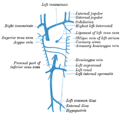 Diagram showing completion of development of the parietal veins.
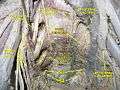 Pelvic contents: male.Superior view.Deep dissection.
References
- Delancey, John O.L. (2016). "73, True pelvis, pelvic floor and perineum". In Standring, Susan (ed.). Gray's Anatomy: The Anatomical Basis of Clinical Practice (41st ed.). Elsevier. pp. 1221–1236. ISBN 978-0-7020-6851-5.
- Sinnatamby, Chummy S. (2011). "5". Last's Anatomy: Regional and Applied (12th ed.). Great Britain: Churchill Livingstone Elsevier. p. 309. ISBN 0-7020-4839-9. Retrieved March 25, 2018.
External links
- Photo of model at Waynesburg College circulation/rightinternaliliacvein
This article incorporates text in the public domain from page 673 of the 20th edition of Gray's Anatomy (1918)
| Authority control |
|---|