Hyperintensity
Hyperintensities refer to areas of high intensity on types of magnetic resonance imaging (MRI) scans of the human brain or that of other mammals that reflect lesions produced largely by demyelination and axonal loss. These small regions of high intensity are observed on T2 weighted MRI images (typically created using 3D FLAIR) within cerebral white matter (white matter lesions, white matter hyperintensities or WMH)[1][2] or subcortical gray matter (gray matter hyperintensities or GMH). They are usually seen in normal aging but also in a number of neurological disorders and psychiatric illnesses. For example, deep white matter hyperintensites are 2.5 to 3 times more likely to occur in bipolar disorder and major depressive disorder than control subjects.[3][4] WMH volume, calculated as a potential diagnostic measure, has been shown to correlate to certain cognitive factors.[5] Hyperintensities appear as "bright signals" (bright areas) on an MRI image and the term "bright signal" is occasionally used as a synonym for a hyperintensity.
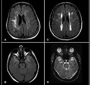
Hyperintensities are commonly divided into 3 types depending on the region of the brain where they are found. Deep white matter hyperintensites occur deep within white matter, periventricular white matter hyperintensities occur adjacent to the lateral ventricles and subcortical hyperintensities occur in the basal ganglia.
Hyperintensities are often seen in auto immune diseases that have effects on the brain.[6]
Postmortem studies combined with MRI suggest that hyperintensities are dilated perivascular spaces, or demyelination caused by reduced local blood flow.[7]
Causes
White matter hyperintensities can be caused by a variety of factors including ischemia, micro-hemorrhages, gliosis, damage to small blood vessel walls, breaches of the barrier between the cerebrospinal fluid and the brain, or loss and deformation of the myelin sheath.[8]
Cognitive effects
In most elderly people, presence of severe WMH and medial temporal lobe atrophy MTA was linked with an increase in frequency of mild cognitive deficits. Studies suggest that a combination of MTA and severe WMH showed more than a fourfold increase in the frequency of mild cognitive deficits.[9] It's also been consistently shown that severe WMH is known to be associated with gait disorders, impaired balance and cognitive disturbances. Certain features of gait pattern associated with WMH are: slight widening of the base, slowing and shortening of stride length and turning en bloc. Speed of cognitive processes and frontal skills may also be impaired in people with WMH.[10][11] Pathological signs of oligodendritic apoptosis and damage to axonal projections have been evident. Sufficient damage to the axons that course through WMH can cause adequate interference with normal neuronal functions.[12]
It is also thought that WMH patients have a negative impact on cognition in Alzheimer's disease population. In Alzheimer's patients, higher WMH are associated with higher amyloid beta deposits, possibly associated with small vessel disease and reduced amyloid beta clearance.[11]
See also
References
- Debette S, Markus HS (2010). "The clinical importance of white matter hyperintensities on brain magnetic resonance imaging: systematic review and meta-analysis". BMJ. 341: c3666. doi:10.1136/bmj.c3666. PMC 2910261. PMID 20660506.
- Habes M, Erus G, Toledo JB, Zhang T, Bryan N, Launer LJ, Rosseel Y, Janowitz D, Doshi J, Van der Auwera S, von Sarnowski B, Hegenscheid K, Hosten N, Homuth G, Völzke H, Schminke U, Hoffmann W, Grabe H, Davatzikos C (2016). "White matter hyperintensities and imaging patterns of brain ageing in the general population". Brain. 139 (Pt 4): 1164–1179. doi:10.1093/brain/aww008. PMC 5006227. PMID 26912649.
- Kempton, Matthew J.; Geddes, JR; Ettinger, U; Williams, SC; Grasby, PM (2008). "Meta-analysis, Database, and Meta-regression of 98 Structural Imaging Studies in Bipolar Disorder". Archives of General Psychiatry. 65 (9): 1017–32. doi:10.1001/archpsyc.65.9.1017. PMID 18762588.
- Videbech, P. (1997). "MRI findings in patients with affective disorder: A meta-analysis". Acta Psychiatrica Scandinavica. 96 (3): 157–68. doi:10.1111/j.1600-0447.1997.tb10146.x. PMID 9296545.
- Brickman, Adam M.; Meier, Irene B.; Korgaonkar, Mayuresh S.; Provenzano, Frank A.; Grieve, Stuart M.; Siedlecki, Karen L.; Wasserman, Ben T.; Williams, Leanne M.; Zimmerman, Molly E. (2012). "Testing the white matter retrogenesis hypothesis of cognitive aging". Neurobiology of Aging. 33 (8): 1699–715. doi:10.1016/j.neurobiolaging.2011.06.001. PMC 3222729. PMID 21783280.
- Theodoridou A, Settas L (2006). "Demyelination in rheumatic diseases". J. Neurol. Neurosurg. Psychiatry. 77 (989): 290–5. doi:10.1136/jnnp.2005.075861. PMC 2077679. PMID 16484634.
- Thomas, Alan J.; Perry, Robert; Barber, Robert; Kalaria, RAJ N.; O'Brien, John T. (2002). "Pathologies and Pathological Mechanisms for White Matter Hyperintensities in Depression". Annals of the New York Academy of Sciences. 977: 333–9. doi:10.1111/j.1749-6632.2002.tb04835.x. PMID 12480770.
- Raz N, Yang Y, Dahle CL, Land S (2012). "Volume of white matter hyperintensities in healthy adults: contribution of age, vascular risk factors, and inflammation-related genetic variants". Biochimica et Biophysica Acta. 1822 (3): 361–369. doi:10.1016/j.bbadis.2011.08.007. PMC 3245802. PMID 21889590.
- Van Der Flier, W M; Van Straaten, EC; Barkhof, F; Ferro, JM; Pantoni, L; Basile, AM; Inzitari, D; Erkinjuntti, T; et al. (2005). "Medial temporal lobe atrophy and white matter hyperintensities are associated with mild cognitive deficits in non-disabled elderly people: The LADIS study". Journal of Neurology, Neurosurgery & Psychiatry. 76 (11): 1497–500. doi:10.1136/jnnp.2005.064998. PMC 1739423. PMID 16227537.
- Gouw, A.A.; Flier, W.M.; Straaten, E.C.W.; Barkhof, F.; Ferro, J.M.; Baezner, H.; Pantoni, L.; Inzitari, D.; et al. (2006). "Simple versus complex assessment of white matter hyperintensities in relation to physical performance and cognition: The LADIS study". Journal of Neurology. 253 (9): 1189–96. doi:10.1007/s00415-006-0193-5. PMID 16998647.
- Birdsill AC, Koscik RL, Jonaitis EM, Johnson SC, Okonkwo OC, Hermann BP, Larue A2, Sager MA, Bendlin BB (2014). "Regional white matter hyperintensities: aging, Alzheimer's disease risk, and cognitive function". Neurobiology of Aging. 35 (4): 769–776. doi:10.1016/j.neurobiolaging.2013.10.072. PMC 3880609. PMID 24199958.CS1 maint: multiple names: authors list (link)
- Bocti, C.; Swartz, R. H.; Gao, F.-Q.; Sahlas, D. J.; Behl, P.; Black, S. E. (2005). "A New Visual Rating Scale to Assess Strategic White Matter Hyperintensities Within Cholinergic Pathways in Dementia". Stroke. 36 (10): 2126–31. doi:10.1161/01.STR.0000183615.07936.b6. PMID 16179569.
External links
- MRI database at www.bipolardatabase.org.