Hemerythrin
Hemerythrin (also spelled haemerythrin; Ancient Greek: αἷμα, romanized: haîma, lit. 'blood', Ancient Greek: ἐρυθρός, romanized: erythrós, lit. 'red') is an oligomeric protein responsible for oxygen (O2) transport in the marine invertebrate phyla of sipunculids, priapulids, brachiopods, and in a single annelid worm genus, Magelona. Myohemerythrin is a monomeric O2-binding protein found in the muscles of marine invertebrates. Hemerythrin and myohemerythrin are essentially colorless when deoxygenated, but turn a violet-pink in the oxygenated state.
Hemerythrin does not, as the name might suggest, contain a heme. The names of the blood oxygen transporters hemoglobin, hemocyanin, hemerythrin, do not refer to the heme group (only found in globins), instead these names are derived from the Greek word for blood. Recent evidence has revealed hemerythrin to be a multi-functional protein – contributing to innate immunity and anterior tissue regeneration in worms.
O2 binding mechanism
The mechanism of dioxygen binding is unusual. Most O2 carriers operate via formation of dioxygen complexes, but hemerythrin holds the O2 as a hydroperoxide (HO2, or -OOH−). The site that binds O2 consists of a pair of iron centres. The iron atoms are bound to the protein through the carboxylate side chains of a glutamate and aspartates as well as through five histidine residues. Hemerythrin and myohemerythrin are often described according to oxidation and ligation states of the iron center:
| Fe2+—OH—Fe2+ | deoxy (reduced) |
| Fe2+—OH—Fe3+ | semi-met |
| Fe3+—O—Fe3+—OOH− | oxy (oxidized) |
| Fe3+—OH—Fe3+— (any other ligand) | met (oxidized) |
The uptake of O2 by hemerythrin is accompanied by two-electron oxidation of the diferrous centre to produce a hydroperoxide (OOH−) complex. The binding of O2 is roughly described in this diagram:
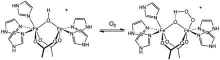 Active site of hemerythrin before and after oxygenation.
Deoxyhemerythrin contains two high-spin ferrous ions bridged by hydroxyl group (A). One iron is hexacoordinate and another is pentacoordinate. A hydroxyl group serves as a bridging ligand but also functions as a proton donor to the O2 substrate. This proton-transfer result in the formation of a single oxygen atom (μ-oxo) bridge in oxy- and methemerythrin. O2 binds to the pentacoordinate Fe2+ centre at the vacant coordination site (B). Then electrons are transferred from the ferrous ions to generate the binuclear ferric (Fe3+,Fe3+) centre with bound peroxide (C).[1][2]
Quaternary structure and cooperativity
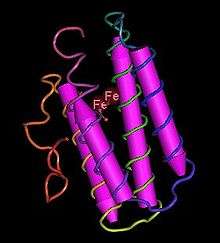
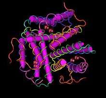
Hemerythrin typically exists as a homooctamer or heterooctamer composed of α- and β-type subunits of 13–14 kDa each, although some species have dimeric, trimeric and tetrameric hemerythrins. Each subunit has a four-α-helix fold binding a binuclear iron centre. Because of its size hemerythrin is usually found in cells or "corpuscles" in the blood rather than free floating.
Unlike hemoglobin, most hemerythrins lack cooperative binding to oxygen, making it roughly 1/4 as efficient as hemoglobin. In some brachiopods though, hemerythrin shows cooperative binding of O2. Cooperative binding is achieved by interactions between subunits: the oxygenation of one subunit increases the affinity of a second unit for oxygen.
Hemerythrin affinity for carbon monoxide (CO) is actually lower than its affinity for O2, unlike hemoglobin which has a very high affinity for CO. Hemerythrin's low affinity for CO poisoning reflects the role of hydrogen-bonding in the binding of O2, a pathway mode that is incompatible with CO complexes which usually do not engage in hydrogen bonding.
Hemerythrin/HHE cation-binding domain
| Hemerythrin HHE cation binding domain | |||||||||
|---|---|---|---|---|---|---|---|---|---|
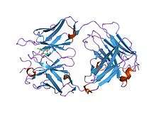 crystal structures of an antibody to a peptide and its complex with peptide antigen at 2.8 angstroms | |||||||||
| Identifiers | |||||||||
| Symbol | Hemerythrin | ||||||||
| Pfam | PF01814 | ||||||||
| InterPro | IPR012312 | ||||||||
| PROSITE | PDOC00476 | ||||||||
| SCOPe | 2hmq / SUPFAM | ||||||||
| |||||||||
The hemerythrin/HHE cation-binding domain occurs as a duplicated domain in hemerythrins, myohemerythrins and related proteins. This domain binds iron in hemerythrin, but can bind other metals in related proteins, such as cadmium in the Nereis diversicolor hemerythrin. It is also found in the NorA protein from Cupriavidus necator, this protein is a regulator of response to nitric oxide, which suggests a different set-up for its metal ligands. A protein from Cryptococcus neoformans (Filobasidiella neoformans) that contains haemerythrin/HHE cation-binding domains is also involved in nitric oxide response.[3] A Staphylococcus aureus protein containing this domain, iron-sulfur cluster repair protein ScdA, has been noted to be important when the organism switches to living in environments with low oxygen concentrations; perhaps this protein acts as an oxygen store or scavenger.[4]
References
- D. M. Kurtz, Jr. "Dioxygen-binding Proteins" in Comprehensive Coordination Chemistry II 2003, Volume 8, Pages 229–260. doi:10.1016/B0-08-043748-6/08171-8
- Friesner, R. A.; Baik, M.-H.; Gherman, B. F.; Guallar, V.; Wirstam, M.; Murphy, R. B.; Lippard, S. J. (2003). "How iron-containing proteins control dioxygen chemistry: a detailed atomic level description via accurate quantum chemical and mixed quantum mechanics/molecular mechanics calculations". Coord. Chem. Rev. 238–239: 267–290. doi:10.1016/S0010-8545(02)00284-9.
- Chow ED, Liu OW, O'Brien S, Madhani HD (September 2007). "Exploration of whole-genome responses of the human AIDS-associated yeast pathogen Cryptococcus neoformans var grubii: nitric oxide stress and body temperature". Curr. Genet. 52 (3–4): 137–48. doi:10.1007/s00294-007-0147-9. PMID 17661046.
- Overton TW, Justino MC, Li Y, Baptista JM, Melo AM, Cole JA, et al. (2008). "Widespread Distribution in Pathogenic Bacteria of Di-Iron Proteins That Repair Oxidative and Nitrosative Damage to Iron-Sulfur Centers". J Bacteriol. 190 (6): 2004–13. doi:10.1128/JB.01733-07. PMC 2258886. PMID 18203837.
Further reading
- Coates, C.J., Decker, H. (2017). "Immunological properties of oxygen transport proteins: hemoglobin, hemocyanin and hemerythrin". Cell. Mol. Life Sci. 74 (2): 293–317. doi:10.1007/s00018-016-2326-7. PMC 5219038. PMID 27518203.CS1 maint: multiple names: authors list (link)
- Karlsen, O.A., Ramsevik, L., Bruseth, L.J., Larsen, Ø., Brenner, A., Berven, F.S., Jensen, H.B. and Lillehaug, J.R. (2005). "Characterization of a prokaryotic haemerythrin from the methanotrophic bacterium Methylococcus capsulatus (Bath)". FEBS J. 272 (10): 2428–2440. doi:10.1111/j.1742-4658.2005.04663.x. PMID 15885093.CS1 maint: multiple names: authors list (link)
- Stenkamp, R.E. (1994). "Dioxygen and hemerythrin". Chem. Rev. 94 (3): 715–726. doi:10.1021/cr00027a008.
External links
- 1HMD - PDB structure of deoxyhemerythrin Themiste dyscrita (sipunculid worm)
- 1HMO – PDB structure of oxyhemerythrin from Themiste dyscrita
- 2MHR – PDB structure of azido-met myohemerythrin from Themiste zostericola (sipunculid worm)
- IPR002063 – InterPro entry for hemerythrin