Bladder exstrophy
Bladder exstrophy is a congenital anomaly that exists along the spectrum of the exstrophy-epispadias complex, and most notably involves protrusion of the urinary bladder through a defect in the abdominal wall. Its presentation is variable, often including abnormalities of the bony pelvis, pelvic floor, and genitalia. The underlying embryologic mechanism leading to bladder exstrophy is unknown, though it is thought to be in part due to failed reinforcement of the cloacal membrane by underlying mesoderm.[1] Exstrophy means the inversion of a hollow organ.[2]
| Bladder exstrophy | |
|---|---|
| Other names | Ectopia vesicae |
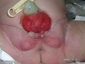 | |
| Female baby with classical bladder exstrophy | |
| Specialty | Medical genetics |
Signs and symptoms
The classic manifestation of bladder exstrophy presents with:
- A defect in the abdominal wall occupied by both the exstrophied bladder as well as a portion of the urethra
- A flattened puborectal sling
- Separation of the pubic symphysis
- Shortening of a pubic rami
- External rotation of the pelvis.
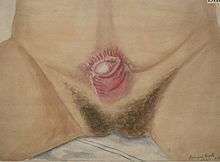
Females frequently have a displaced and narrowed vaginal orifice, a bifid clitoris, and divergent labia.[3]
Cause
The cause is not yet clinically established but is thought to be in part due to failed reinforcement of the cloacal membrane by underlying mesoderm.
Diagnosis
In a small retrospective study of 25 pregnancies five factors were found to be strongly associated with a prenatal diagnosis of bladder exstrophy:
- Inability to visualize the bladder on ultrasound
- A lower abdominal bulge
- A small penis with anteriorly displaced scrotum
- A low set umbilical insertion
- Abnormal widening of the iliac crests
While a diagnosis of bladder exstrophy was made retrospectively in a majority of pregnancies, in only three cases was a prenatal diagnosis made.[4]
Management
The extreme rarity of the disease limits the surgical opportunities to practice the complex closure required in these patients. For this reason, patients have the best outcomes when the bladder closures are performed at high volume centers where surgical and nursing teams have extensive experience in caring for the disease. [5] The highest volume center in the United States, and the world, is the Johns Hopkins Hospital in Baltimore, Maryland; they have seen over 1300 exstrophy patients in the past 50 years. [6]
Upon delivery, the exposed bladder is irrigated and a non-adherent film is placed to prevent as much contact with the external environment as possible. In the event the child was not born at a medical center with an appropriate exstrophy support team then transfer will likely follow. Upon transfer, or for those infants born at a medical center able to care for bladder exstrophy, imaging may take place in the first few hours of life prior to the child undergoing surgery.[3]
Primary (immediate) closure is indicated only in those patients with a bladder of appropriate size, elasticity, and contractility as those patients are most likely to develop a bladder of adequate capacity after early surgical intervention.[7]
Conditions that are absolute contraindications despite bladder adequacy include duplication of the penis or scrotum and significant bilateral hydronephrosis.
Surgery
Modern therapy is aimed at surgical reconstruction of the bladder and genitalia. Both males and females are born with this anomaly. Treatment is similar.
In males treatments have been: In the modern staged repair of exstrophy (MSRE) the initial step is closure of the abdominal wall, often requiring a pelvic osteotomy. This leaves the patient with penile epispadias and urinary incontinence. At approximately 2–3 years of age the patient then undergoes repair of the epispadias after testosterone stimulation. Finally, bladder neck repair usually occurs around the age of 4–5 years, though this is dependent upon a bladder with adequate capacity and, most importantly, an indication that the child is interested in becoming continent. In some of the bladder reconstructions, the bladder is augmented with the addition of a segment of the large intestines to increase the volume capacity of the reconstructed bladder. (http://www.med.umich.edu/1libr/urology/BladderAugmentation.pdf) In the complete primary repair of exstrophy (CPRE) the bladder closure is combined with an epispadias repair, in an effort to decrease costs and morbidity.[8] This technique has, however, led to significant loss of penile and corporal tissue, particularly in younger patients.[9]
In females treatment has included: Surgical reconstruction of the clitoris which is separated into two distinct bodies. Surgical reconstruction to correct the split of the mons, redefine the structure of the bladder neck and urethra. Vaginoplasty will correct the anteriorly displaced vagina. If the anus is involved, it is also repaired. Fertility remains and women who were born with bladder extrophy usually develop prolapse due to the weaker muscles of the pelvic floor.[10]
Prognosis
The most important criterion for improving long-term prognosis is success of the initial closure.[11][12] If a patient requires more than one closure their chance of continence drops off precipitously with each additional closure - at just two closures the chance of voiding continence is just 17%.[13]
Even with successful surgery, people may have long-term complications.[14] Some of the most common include:
Epidemiology
Occurring at a rate between 1 in 10,000 to 1 in 50,000 [15] with a male-to-female ratio of 2.3-6:1,[16][17][18] bladder exstrophy is relatively rare. For those individuals with bladder exstrophy who maintain their ability to reproduce, the risk of bladder exstrophy in their children is approximately 500-fold greater than the general population.[16]
References
- Muecke EC: The role of the cloacal membrane in exstrophy: the first successful experimental study. J Urol 1964; 92:659.
- Larsen, WJ. (2001). Human embryology (3 ed.). Philadelphia, Pa.: Churchill Livingstone. p. 275. ISBN 0-443-06583-7.
- Gearhart JP, Mathews R. Exstrophy-epispadias complex. In: Wein AJ, ed. Campbell-Walsh Urology. 10th ed. Philadelphia, Pa: Saunders Elsevier; 2011:chap 124.
- Gearhart JP, Ben-Chaim J, Jeffs RD, et al: Criteria for the prenatal diagnosis of classic bladder exstrophy. Obstet Gynecol 1995; 85:961.
- Nelson CP, Dunn RL, Wei JT, Gearhart JP. Surgical repair of bladder exstrophy in the modern era: contemporary practice patterns and the role of hospital case volume. Journal of Urology. 2005 Sep;174(3):1099-102.
- Kasprenski, M., Benz, K.S., Maruf, M., Jayman, J., DiCarlo, H., Gearhart, J.P. Modern Management of the Failed Bladder Exstrophy Closure: A 50 Year Experience. European Urology Focus. 2018.
- Gearhart JP, Jeffs RD: The bladder exstrophy-epispadias complex. In: Walsh PC, et al ed. Campbell's urology, 7th ed. Philadelphia: WB Saunders; 1998:1939.
- Grady, Mitchell, 1999. Grady R, Mitchell ME: Complete repair of exstrophy. J Urol 1999; 162:1415.
- Husmann, Gearhart, 2004. Husmann DA, Gearhart JP: Loss of penile glans and/or corpora following primary repair of bladder exstrophy using the complete penile disassembly technique. J Urol 2004; 172:1696.
- Moliterno, David (2013). Therapeutic advances in thrombosis. Chichester, West Sussex: Wiley-Blackwell. pp. 779–798. ISBN 9781405196253.
- The importance of a successful initial bladder closure in the surgical management of classical bladder exstrophy: analysis of 144 patients treated at the Johns Hopkins Hospital between 1975 and 1985. 1987 Feb;137(2):258–62.
- Closure of the exstrophic bladder: an evaluation of the factors leading to its success and its importance on urinary continence. 1989 Aug;142(2 Pt 2):522–4–discussion542–3.
- Failed exstrophy closure: management and outcome. 2010 Aug;6(4):381–4.
- Gargollo PC, Borer JG (2007). "Contemporary outcomes in bladder exstrophy". Current Opinion in Urology. 17 (4): 272–80. doi:10.1097/MOU.0b013e3281ddb32f. PMID 17558272.
- Lattimer JK, Smith MJK: Exstrophy closure: a follow up on 70 cases. J Urol 1966; 95:356.
- Shapiro E, Jeffs RD, Gearhart JP, Lepor H: Muscarinic cholinergic receptors in bladder exstrophy: insights into surgical management. J Urol 1985; 134:309.
- Ives E, Coffey R, Carter CO: A family study of bladder exstrophy. J Med Genet 1980; 17:139.
- Lancaster PAL: Epidemiology of bladder exstrophy: a communication from the International Clearinghouse for Birth Defects monitoring systems. Teratology 1987; 36:221.
External links
| Classification | |
|---|---|
| External resources |
| Wikimedia Commons has media related to Bladder exstrophy. |