Bilophila wadsworthia
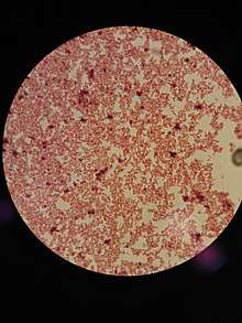
Bilophila wadsworthia is a gram-negative, obligately anaerobic, catalase-positive, bile-resistant, and asaccharolytic bacillus. This bacterium is about 0.7 μm wide by 1.0–10.0 μm long in colony and the cells are pleomorphic with irregular cell walls.[1][2] This bacterial species is mostly urease positive; around 75% of the strains are urease positive.[3] Less than 0.01% of the human gut microbiota is this bacterium.[3] B. wadsworthia is linked to various diseases and is not well known due to frequent misidentification of the bacteria. The two unique characteristics of B. wadsworthia are the production of hydrogen sulfide and the rapid catalase reaction. This bacterium is vulnerable to the antibiotics: imipenem, ceoxitin, and ticarcillin, which are all β-lactam antibiotics.
| Bilophila wadsworthia | |
|---|---|
| Scientific classification | |
| Kingdom: | Bacteria |
| Phylum: | |
| Order: | |
| Family: | |
| Genus: | |
| Species: | B. wadsworthia |
| Binomial name | |
| Bilophila wadsworthia Baron, 1990 | |
Biology
Type and morphology
B. wadsworthia is a gram-negative, catalase-positive, and usually urease-positive bacterium.[3] Although most strains are urease positive, there are some strains that are urease negative. This bacterium, due to its slow nature to grow in standard media for anaerobes, is often misidentified as other anaerobes or not identified at all.[3] The best identifier for this bacterium is a transparent colony with a black center in BBE agar; the black center is ferrous sulfide, which is created by the hydrogen sulfide the bacteria produces.[3][2] Another unique characteristic of B. wadsworthia is its positive catalase reaction with a 15% hydrogen peroxide reagent; its catalase reaction is unlike other catalase-positive species because the reaction is explosive with bubble formation and is very quick.[4] The most accurate method to identify this bacterium from other similar species is through gas liquid chromatography (GLC); GLC results show that there are a lot of acetic acid peaks and minimal succinic acid present. B. wadsworthia is non-motile and is non-spore forming.[1] In addition, this bacterium has irregular cell walls and no flagella attached to it. The two factors that stimulate its growth are bile and pyruvate; specifically, 20% bile and 1% pyruvate.[2]
Metabolism
One of the main processes that occur in B. wadsworthia is hydrogen sulfide production, which is the product responsible for the bacterium's signature black dot.[5] B. wadsworthia is able to produce hydrogen sulfide through its taurine desulfonation pathway using isethionate sulfite-lyase (IslA).[5] The bacterium converts taurine to hydrogen sulfide when it respires taurine. The production of hydrogen sulfide is connected to the human intestinal microbiota; although there are some benefits to hydrogen sulfide production in the gut like cardioprotection, hydrogen sulfide production also contributes to disease pathology. Production of hydrogen sulfide has been linked to irritable bowel disease (IBD) by damaging the gut epithelium's mucus layer and to colorectal cancer.[5] In addition, during treatments with antibiotics, hydrogen sulfide can aide opportunistic bacteria grow leading to antibiotic resistance.[5] Future research on controlling hydrogen sulfide production may help address B. wadsworthia's contribution to diseases.
Culture growth on bacteriodes Bile esculin (BBE) agar
The bacteria must be grown on this agar for at least 3 days in order to see colony formation. Starting from day 3, two possible types of colonies appear. First, a 1-2 mm diameter convex and irregular colony with a black center is visible.[4] Second, a translucent umbonate and circular colony with a dark center is present. In addition, β-lactamase is not produced in this agar and the colonies formed in BBE agar pass the positive urease test.[1] BBE agar is the optimal agar for B. wadsworthia colony growth.
Culture growth on Brucella agar
The bacteria similar to the BBE agar must be incubated on the agar for at least 4 days. In this agar, a 0.6-0.8 mm diameter raised, circular erose colony is visible.[2] Also, the colony is gray and translucent.[3]
Location
Although the preferred location for this bacterium is still unknown, it is mostly found in the lower parts of the gastrointestinal tract.[3] This bacterium is considered virulent in nature because it is commonly found in patients with appendicitis, gangrenous appendicitis, and the blood cultures of patients with liver abscesses; it is the third-most abundant anaerobic bacterium found in patients with appendicitis.[3][2][4] Also, they are found in the feces samples of healthy patients.[3][2] In non-human cases, this bacterium is found in dogs with periodontal disease.[3] In rare cases, this bacterium may be found in saliva and vaginal samples.[3][2]
Other specimen locations where B. wadsworthia have been found include:
- Scrotal abscess
- Mandibular osteomyelitis[6][4]
- Axillary Hidradenitis suppurativa[7][4]
- Sepsis
- Cholecystitis[3]
- Bartholinitis[3]
Discovery
B. wadsworthia was first identified in 1988 by E. J. Baron,[8] a retired pathology professor from Stanford University, in specimens collected from patients with perforated appendicitis and gangrenous appendicitis; the bacterium was also found in healthy fecal specimens.[3] This bacterium was categorized into the genus Bilophila because of its bile-loving and growing nature.[4] Baron named the species wadsworthia after the Wadsworth Anaerobe Laboratory,[9] which was where the bacteria was first identified.
References
- Kasten, M J; Rosenblatt, J E; Gustafson, D R (September 1992). "Bilophila wadsworthia bacteremia in two patients with hepatic abscesses". Journal of Clinical Microbiology. 30 (9): 2502–2503. ISSN 0095-1137. PMC 265535. PMID 1401025.
- Baron, E. J.; Summanen, P.; Downes, J.; Roberts, M. C.; Wexler, H.; Finegold, S. M. (1989-12-01). "Bilophila wadsworthia, gen. nov. and sp. nov., a Unique Gram-negative Anaerobic Rod Recovered from Appendicitis Specimens and Human Faeces". Microbiology. 135 (12): 3405–3411. doi:10.1099/00221287-135-12-3405. ISSN 1350-0872. PMID 2636263.
- Baron, Ellen Jo (April 1997). "Bilophila wadsworthia: a Unique Gram-negative Anaerobic Rod". Anaerobe. 3 (2–3): 83–86. doi:10.1006/anae.1997.0075. ISSN 1075-9964. PMID 16887567.
- Summanen, P. H.; Jousimies-Somer, H.; Manley, S.; Bruckner, D.; Marina, M.; Goldstein, E. J. C.; Finegold, S. M. (1995-06-01). "Bilophila wadsworthia Isolates from Clinical Specimens". Clinical Infectious Diseases. 20 (Supplement_2): S210–S211. doi:10.1093/clinids/20.supplement_2.s210. ISSN 1537-6591. PMC 265400. PMID 1629348.
- Peck, Spencer C.; Denger, Karin; Burrichter, Anna; Irwin, Stephania M.; Balskus, Emily P.; Schleheck, David (2019-02-04). "A glycyl radical enzyme enables hydrogen sulfide production by the human intestinal bacterium Bilophila wadsworthia". Proceedings of the National Academy of Sciences. 116 (8): 3171–3176. doi:10.1073/pnas.1815661116. ISSN 0027-8424. PMC 6386719. PMID 30718429.
- "Osteomyelitis - Symptoms and causes". Mayo Clinic. Retrieved 24 March 2019.
- "Hidradenitis suppurativa - Symptoms and causes". Mayo Clinic. Retrieved 24 March 2019.
- "Ellen Baron's Profile - Stanford Profiles". profiles.stanford.edu. Retrieved 24 March 2019.
- "Laboratory Services". New York State Department of Health, Wadsworth Center. 5 June 2015. Retrieved 24 March 2019.