We need you! Join our contributor community and become a WikEM editor through our open and transparent promotion process.
Ultrasound: Soft tissue
From WikEM
Contents
Background
- Soft tissue ultrasound can help with differentiating abscess from cellulitis
- Many types of foreign bodies can be visualized
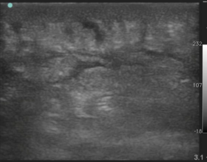
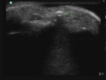
Cellulitis
Images
Instructions
- Select linear probe (high frequency probe)
- Scan area of concern (orientation of probe not as important)
- Rotate 90° over area of concern
Findings
- Positive
- Cobblestoning - thin lines of fluid between fat globules
- Loss of tissue plain definition
Pearls and Pitfalls
- Cobblestone can also be present in:
- Lymphedema
- Pitting edema secondary to HF
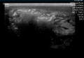
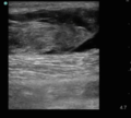
Abscess
Novice sonographers can predict a positive I&D with SN 0.97 and SP 0.67 (vs clinical exam 0.76 and 0.83)[1]
Images
Gas gangrene
Abscess
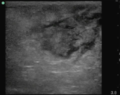
Hematoma
Hematoma
Instructions
- Select linear probe (high frequency probe)
- Scan area of concern (orientation of probe not as important)
- Rotate 90° over area of concern
- If hypoechoic area is identified, apply gentle pressure over area
Findings
- Positive scan (not all elements are required to make a diagnosis)
- Fluid collect seen has heteroechoic or hypoechoic circular area
- Hyperechoic ring
- Posterior acoustic enhancement
- Swirling or Squish Sign (movement of abscess debris) with compression
Pearls and Pitfalls
- Color flow may be used to differentiate vascular and lymphatic structures
- In the inguinal crease strangulated bowel can mimic abscess
- The collection seen under U/S may not correspond with actual collection size
- Hematomas can look similar to abscess, so the right clinical context is needed
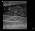
Foreign Body
Images
Foreign body in finger
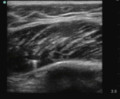
Needle tip in muscle
Instructions
- Select linear probe (high frequency probe)
- Scan area of concern (orientation of probe not as important)
- Rotate 90° over area of concern
Findings
- FB can show 2 different signs
- Acoustic shadow - Ring down appearance
- Common with wood and splinters
- Reverberation
- Common with metal such are retained insulin needle
- Acoustic shadow - Ring down appearance
Pearls and Pitfalls
- U/S is no sensitive for FB (U/S will miss a substantial amount of FBs)
- Xray or other modality may be needed for look during negative exams
- Real-time U/S can aid in FB removal
- Water baths may be helpful for extremities
- Scar tissue may mimic FB
See Also
External Links
References
- ↑ Berger, T, et al. Bedside ultrasound performed by novices for the detection of abscess in ED patients with soft tissue infections. Am J Emerg Med. 2012; 30(8):1569-73. doi: 10.1016/j.ajem.2011.08.002.