We need you! Join our contributor community and become a WikEM editor through our open and transparent promotion process.
Brain MRI
From WikEM
(Redirected from MRI Brain)
Contents
Background
- MRI uses magnetic fields and radiowaves to develop high definition imaging of the brain and excellent tissue contrast
- No radiation associated with imaging
- Ideal for looking at brain parenchyma and midbrain
- Contrast is commented on by signal intensity
- Dark areas are hypointense
- Bright areas are hyperintense
Ordering Studies
MR Imaging (for Rule-Out CVA or TIA)
- MRI Brain with DWI (without contrast) AND
- Cervical vascular imaging (ACEP Level B in patients with high short-term risk for stroke):[1]
- MRA brain (without contrast) AND
- MRA neck (without contrast)
- May instead use Carotid CTA or US (Carotid US slightly less sensitive than MRA)[2] (ACEP Level C)
Contrast only needed if concern for malignancy/mass
MRI Modalities
T1 Weighted Imaging
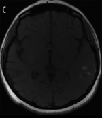
- Ideal for brain parenchyma
- With the addition of contrast, this can differentiate causes of inflammation
- Fluid is hypointense (similar to CT imaging)
- Methemoglobin, fat, and protein are hyperintense
T2 Weighted Imaging
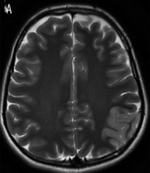
- Highlights CSF
- Good for identifying tissue edema around pathologic areas
- Fluid is hyperintense (reverse of T1)
- Tissue tends to be more hypointense
Fluid Attenuation Inversion Recovery (FLAIR)
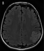
- Appears as T2 images with hypointense CSF- cancels out CSF so you can differentiate CSF from other fluid
- Ideal for identifying tumors/GBS
- Also used to identify leptomeningeal enhancement in meningitis
Diffusion Weighted Imaging (DWI)
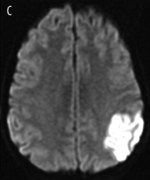
- A method of measuring the Brownian motion of water molecules
- Diffusion within the intracellular fluid, diffusion within extracellular fluid, and between these areas will differ depending on pathology
- Ideal for cellular swelling especially in acute ischemic stroke which will be hyperintense
Blood
| Age of Blood | T1 Imaging | T2 Imaging |
|---|---|---|
| Hyperacute | Iso | Bright |
| Acute | Iso/Dark | Dark |
| 1-3 Days | Bright | Dark |
| 1-2 Wks | Bright | Bright |
| 2-3 Wks | Iso/Dark | Dark |
See Also
References
- ↑ ACEP Clinical Policy: Suspected Transient Ischemic Attackfull text
- ↑ Nederkoorn PJ, Mali WP, Eikelboom BC, et al. Preoperative diagnosis of carotid artery stenosis. Accuracy of noninvasive testing. Stroke. 2002;33:2003-2008.