We need you! Join our contributor community and become a WikEM editor through our open and transparent promotion process.
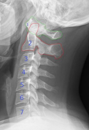
C-Spine X-Ray
From WikEM
Contents
Background
- Make sure that the C7-T1 junction is adequately visualized
- Obtain swimmer's view or oblique view if inadequate
- Peds
- Most pediatric fractures occur higher than C3
- Pseudosubluxation of C2-C3 is common in children <8yr
- To distinguish from true dislocation or fracture:
- Draw line from cortex of post arch of C1 to cortex of posterior arch of C3
- This line should pass through or be <1mm ant to posterior arch of C2
- To distinguish from true dislocation or fracture:
- Most common approach is to evaluate three parallel vertical columns
- Anterior column: alternating vertebral bodies and intervertebral disks surrouded by anulus fibrosus and anterior longitudinal ligament
- Middle column: posterior parts of annulus fibrosis and posterior vertebral wall, posterior lognitudinal ligament, spinal cord, paired laminae and pedicles, articulating facets, transverse processes, nerve roots and vertebral arteries/veins
- Posterior column: spinous process, nuchal ligament, interpsinous and supraspinous ligaments, and ligamentum flavum.
- Disruption of one column is generally stable. Disruption of two or more is unstable.
Measurements (Normal)
- Predental space (anterior aspect of odontoid to post aspect of ant ring of C1)
- Adult <3mm
- Peds <5mm
- Widening of space suggests Jefferson burst fracture of C1
- Anterior soft tissue
- Distance between ant border of C2 and post pharynx should be <6mm in adults and peds
- Distance between ant border of C6 and post trachea should be <22 mm in adults
- Should be <14mm in children <15yr or less than width of vertebral body at each level
- Bones
- Vertebral body: Anterior height should be no more than 3mm shorter than posterior height
Lateral View
- Alignment
- Disruption in the anterior, posterior, or spinolaminal lines
- Bones
- Obvious fracture
- Disruption of ring of C1
- Double facet sign indicates fractured articular facet
- Loss of vertebral height
- Cartilage
- Intervertebral disc space height and length should be uniform
- Narrowing: disc herniation or adjacent vertebral fracture
- Widening: posterior ligamentous injury
- Intervertebral disc space height and length should be uniform
- Soft tissue
- Widening of the prevertebral soft tissue suggests fracture
AP View
- Alignment of spinous processes
- Distance between spinous processes
- Uniformity and height of vertebrae
Odontoid View
- Spacing of dens and lateral masses
- Lateral alignment of C1 and C2
- Uniformity of bones
X-ray vs CT
- Plain radiographs may be appropriate in low-risk patients
- High risk patients requiring CT:
- Closed head injury
- Neurologic deficits
- High energy trauma
- Unreliable examination
- Pain out of proportion to exam
- Inadequate plain films