We need you! Join our contributor community and become a WikEM editor through our open and transparent promotion process.
Bladder ultrasound
From WikEM
Contents
Background
- Bladder ultrasounds can be used independently for volume measurements or in conjunction with other exams such as FAST, renal studies, and pelvic ultrasounds
- Bladder volume = length x width x height x 0.52[1]
Indications
- Urinary retention
- Urinary catheter confirmation
- Free fluid in the pelvis
- Determine post void residual
- Nephrolithiasis in the UVJ
- Typically included in renal ultrasound and FAST exams
Technique
- Select probe
- Phased array or curvilinear probe
- Location
- Suprapubic
- Landmarks
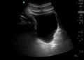
- Identification: anechoic (black) fluid within a structure defined by hyperechoic (white) appearing borders
- Obtain sagittal and transverse images
- Use calc function to attain bladder volume
- Optimize image quality
- May need to turn down far gain secondary to acoustic enhancement from bladder
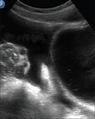
- May use power Doppler over the inferior border can demonstrate ureteral jets
Findings
- Bladder volume/post-void residual
- Use calc mode and measure in 3 dimensions (anterior posterior, right left, and superior inferior)
- Stones
- Ureteral jets can indicate patent ureter
- Bladder bulge may indicated a UVJ stone[2]
- Twinkle Sign: Rapid alternation of color immediately behind a stationary echogenic object, acquiring a false appearance of movement in color Doppler mode
- Free fluid
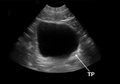
- Anechoic (black) fluid outside the bladder is suggestive of free fluid
- Look for 'pointy edges' which increases suspicion for fluid not in another structure (cyst, ovary, bowel, etc)
Images
Normal

Ureteral jet
Abnormal
Free fluid superior to the bladder
Right UVJ stone
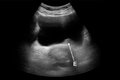
Right UVJ stone
Pearls and Pitfalls
- Ureteral jets indicate patent ureter but the absence of it does not mean obstruction
Documentation
A bedside ultrasound was conducted to assess for bladder volume with clinical indications of urinary retention. The bladder was identified and viewed in the transverse and sagittal plane. The bladder volume was calculated to be ***ml.
See Renal ultrasound, FAST exam, and Transabdominal pelvic ultrasound for further documentation for in other indications
Clips
Normal Exam


External Links
See Also
- Ultrasound (main)
- Ultrasound: In Shock and Hypotension
- Ultrasound: Pelvic
- Renal ultrasound
- Ultrasound: Signs
References
- ↑ Dicuio M et al. Measurements of urinary bladder volume: comparison of five ultrasound calculation methods in volunteers. Arch Ital Urol Androl. 2005 Mar;77(1):60-2.
- ↑ Bomann JS, Seman M, Sutijono D, et al. Bladder bulge: unifying old and new sonographic bladder wall abnormalities in ureterolithiasis. West J Emerg Med. 2012;13(6):517-523.