We need you! Join our contributor community and become a WikEM editor through our open and transparent promotion process.
Difference between revisions of "Tibial plateau fracture"
From WikEM
(→Imaging) |
|||
| (2 intermediate revisions by one other user not shown) | |||
| Line 16: | Line 16: | ||
==Evaluation== | ==Evaluation== | ||
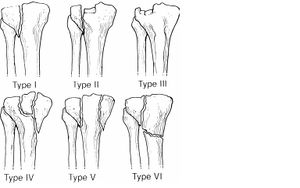
| + | [[File:Schatzker Classification.jpg|thumb|Schatzker Classification of Tibial Plateau Fractures]] | ||
===Imaging=== | ===Imaging=== | ||
*AP, lateral, oblique views (internal for lateral plateau, external for medial plateau). Tunnel view may also be helpful. | *AP, lateral, oblique views (internal for lateral plateau, external for medial plateau). Tunnel view may also be helpful. | ||
| Line 22: | Line 23: | ||
===Schatzker Classification=== | ===Schatzker Classification=== | ||
| − | |||
*Schatzker I Lateral split | *Schatzker I Lateral split | ||
*Schatzker II Split with depression | *Schatzker II Split with depression | ||
| Line 32: | Line 32: | ||
==Management== | ==Management== | ||
*Knee immobilizer with non-weightbearing and ortho referral in 2-7d | *Knee immobilizer with non-weightbearing and ortho referral in 2-7d | ||
| + | *Emergent surgical management if open or if neurovascular compromise | ||
==Disposition== | ==Disposition== | ||
| Line 37: | Line 38: | ||
**Significant displacement or depression | **Significant displacement or depression | ||
**Suspected or documented ligamentous injury | **Suspected or documented ligamentous injury | ||
| + | *Indications for surgery | ||
| + | **Articular stepoff > 3mm | ||
| + | **Condylar widening > 5mm | ||
| + | **Varus/valgus instability | ||
| + | **All medial plateau fractures | ||
| + | **All bicondylar fractures | ||
==See Also== | ==See Also== | ||
| Line 43: | Line 50: | ||
==References== | ==References== | ||
| + | <references/> | ||
[[Category:Orthopedics]] | [[Category:Orthopedics]] | ||
Latest revision as of 21:01, 9 May 2017
Contents
Background
- ACL and MCL injuries associated with lateral plateau fracture
- PCL and LCL associated with medial plateau fracture
- Compartment syndrome may occur
- Segond Fracture
- Avulsion fracture of margin of lateral tibial plateau just below joint line
- Associated with tear of ACL and meniscal ligaments
Clinical Features
- Occurs via axial load that drives femoral condyle into tibia
Differential Diagnosis
Knee diagnoses
Acute Injury
- Knee fractures
- Patella fracture
- Tibial plateau fracture
- Knee dislocation
- Patella dislocation
- Segond fracture
- Meniscus and ligament knee injuries
- Patellar Tendinitis (Jumper's knee)
- Patellar tendon rupture
- Quadriceps tendon rupture
Nontraumatic/Subacute
- Septic Joint
- Gout
- Popliteal cyst (Baker's)
- Prepatellar bursitis (nonseptic)
- Septic bursitis
- Pes anserine bursitis
- Patellofemoral syndrome (Runner's Knee)
- Patellar Tendinitis (Jumper's knee)
- Osgood-Schlatter disease
- Arthritis
Distal Leg Fractures
- Tibial plateau fracture
- Tibial shaft fracture
- Pilon fracture
- Maisonneuve fracture
- Tibia fracture (peds)
- Ankle fracture
- Foot and toe fractures
Evaluation
Imaging
- AP, lateral, oblique views (internal for lateral plateau, external for medial plateau). Tunnel view may also be helpful.
- AP - line drawn at lateral margin of femur should not have >5mm of tibia beyond it
- CT or MRI should be considered if plain film negative but high clinical suspicion based on mechanism or inability to bear weight
Schatzker Classification
- Schatzker I Lateral split
- Schatzker II Split with depression
- Schatzker III Pure lateral depression
- Schatzker IV Pure medial depression
- Schatzker V Bicondylar
- Schatzker VI Split extends to metadiaphysis
Management
- Knee immobilizer with non-weightbearing and ortho referral in 2-7d
- Emergent surgical management if open or if neurovascular compromise
Disposition
- Indications for referral within 48hr:
- Significant displacement or depression
- Suspected or documented ligamentous injury
- Indications for surgery
- Articular stepoff > 3mm
- Condylar widening > 5mm
- Varus/valgus instability
- All medial plateau fractures
- All bicondylar fractures
See Also
References
Authors
Ross Donaldson, Jay, Michael Holtz, Daniel Ostermayer, Neil Young

