

Disease Specifics
 ShareCompartir
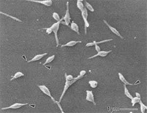
Electron micrograph of Mycoplasma pneumoniae cells. The arrows indicate the attachment organelles.
Waites KB, Talkington DF. Mycoplasma pneumoniae and its role as a human pathogen. Clin Microbiol Rev. 2004;17:697–728. doi: 10.1128/CMR.17.4.697-728.2004
Mycoplasma pneumoniae infection is a type of atypical bacterial pneumonia that is characterized by a relatively long incubation period (approximately 1–4 weeks) and a wide spectrum of clinical symptoms and disease manifestations. While the true magnitude of this health issue in the United States remains unknown, an estimated 2 million cases of M. pneumoniae infection occur each year. It is also estimated that M. pneumoniae infection accounts for between 1 and 10 out of every 50 cases of community-acquired pneumonia.
Pneumonia resulting from an M. pneumoniae infection was originally differentiated from pneumococcal pneumonia because some cases failed to respond to antibiotic treatment with sulfonamides or penicillin (beta-lactams). The unresponsiveness of M. pneumoniae to beta-lactam antibiotics was deemed “atypical.”
The varied presentation and limited diagnostic methods available present unique challenges for accurately identifying M. pneumoniae cases and appropriately treating patients.
Mycoplasma pneumoniae
M. pneumoniae was first isolated from the sputum of a patient with primary “atypical” pneumonia in 1944. The bacterium was considered to be a virus until it was observed that antibiotics could be effective against it. M. pneumoniae is a very small bacterium that thrives within osmotically stable environments, such as the human host.
The cell volume of M. pneumoniae is less than 5% of the cell volume of a typical bacillus, allowing it to pass through filters that are commonly used to remove bacteria. This small cellular mass also means that it cannot be detected by light microscopy and it does not produce visible turbidity in liquid growth media. Specialized media are used to grow M. pneumoniae that allows for a visual confirmation of a positive culture.
M. pneumoniae lacks a rigid cell wall, allowing it to alter its size and shape to suit its surrounding conditions. It is also intrinsically resistant to antimicrobials, like beta-lactams, that work by targeting the cell wall. Due to its lack of a cell wall, M. pneumoniae is extremely susceptible to desiccation; therefore, transmission of infection from person-to-person by airborne droplets only occurs through close contact.
Pathogenesis
M. pneumoniae is transmitted through airborne droplets from person-to-person and is exclusively a human pathogen. M. pneumoniae is primarily an extracellular pathogen that has evolved a specialized attachment organelle for close association with host cells. M. pneumoniae’s attachment is paramount to the survival and existence of the bacterium within the host. The close association between M. pneumoniae and the host cells prevents the bacterium from being removed by the host’s mucociliary clearance mechanisms. The bacterium attaches to and damages the respiratory epithelial cells at the base of cilia, activating the innate immune response and producing local cytotoxic effects.
M. pneumoniae produces a unique virulence factor known as Community Acquired Respiratory Distress Syndrome (CARDS) toxin. The CARDS toxin most likely aids in the colonization and pathogenic pathways of M. pneumoniae, leading to inflammation and airway dysfunction. While M. pneumoniae primarily lives on the surface of the respiratory epithelial cells, it has also been shown to invade tissues and replicate intracellularly. The endocytosis of M. pneumoniae by the host cells could aid in the establishment of a latent or chronic disease state, facilitate the bacterium in evading an immune response, or interfere with the efficacy of certain drug therapies.
References
- Marston BJ, Plouffe JF, File Jr TM, Hackman BA, Salstrom SJ, Lipman HB, Kolczak MS, Breiman RF. Incidence of community-acquired pneumonia requiring hospitalization. Results of a population-based active surveillance study in Ohio. The Community-Based Pneumoniae Incidence Study Group. Arch Intern Med. 1997;157:1709–18.
- Kannan TR, Baseman JB. ADP-ribosylating and vacuolating cytotoxin of Mycoplasma pneumoniae represents unique virulence determinant among bacterial pathogens. Proc Natl Acad Sci USA. 2006;103:6724–9.
- Porath A, Schlaeffer F, Lieberman D. The epidemiology of community-acquired pneumonia among hospitalized adults. J Infect. 1997;34:41–8.
- Waites KB, Atkinson TP. The role of mycoplasma in upper respiratory infections. Curr Infect Dis Rep. 2009;11:198–206.
- Waites KB, Talkington DF. Mycoplasma pneumoniae and its role as a human pathogen. Clin Microbiol Rev. 2004;17:697–728.
- Page last reviewed: March 15, 2016
- Page last updated: March 15, 2016
- Content source: