

Diagnosis
 ShareCompartir
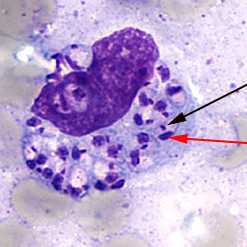
Light-microscopic examination of a stained bone marrow specimen from a patient with visceral leishmaniasis—showing a macrophage (a special type of white blood cell) containing multiple Leishmania amastigotes (the tissue stage of the parasite). Note that each amastigote has a nucleus (red arrow) and a rod-shaped kinetoplast (black arrow). Visualization of the kinetoplast is important for diagnostic purposes, to be confident the patient has leishmaniasis. (Credit: CDC/DPDx)
Various laboratory methods can be used to diagnose leishmaniasis—to detect the parasite as well as to identify the Leishmania species (type). Some of the methods are available only in reference laboratories. In the United States, CDC staff can assist with the testing for leishmaniasis.
Tissue specimens—such as from skin sores (for cutaneous leishmaniasis) or from bone marrow (for visceral leishmaniasis)—can be examined for the parasite under a microscope, in special cultures, and in other ways. Blood tests that detect antibody (an immune response) to the parasite can be helpful for cases of visceral leishmaniasis; tests to look for the parasite itself usually also are done.
- Page last reviewed: January 10, 2013
- Page last updated: January 10, 2013
- Content source: