

Chapter 22 - Evaluation of predictive genetic tests for common diseases: bridging epidemiological, clinical, and public health measures
 ShareCompartir
This website is archived for historical purposes and is no longer being maintained or updated.
Human Genome Epidemiology (2nd ed.): Building the evidence for using genetic information to improve health and prevent disease
“The findings and conclusions in this book are those of the author(s) and do not
necessarily represent the views of the funding agency.”
necessarily represent the views of the funding agency.”
These chapters were published with modifications by Oxford University Press (2010)
A. Cecile J. W. Janssens, Marta Gwinn, and Muin J. Khoury
Figures 22-1
Disease risks of carriers and noncarriers in genetic testing for monogenic disorders (10). The genetic variants tested are CAG repeats in 4p16.3 for Huntington disease (7), BRCA1/BRCA2 mutations for breast cancer (8), and MLH1/hMSH2 mutations for colorectal cancer (9).
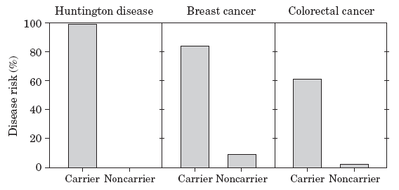
[A text description of this chart is also available.]
Figure 22-2
Disease risks of carriers and noncarriers in single genetic testing for multifactorial disorders (10). Data on the odds ratios of the genetic variants are obtained from the literature (PPARG (11), CAPN10 (12), TCF7L2 (13)). For the calculation of the disease risks from the published odds ratios, we assumed a lifetime risk of type 2 diabetes of 33% (14).
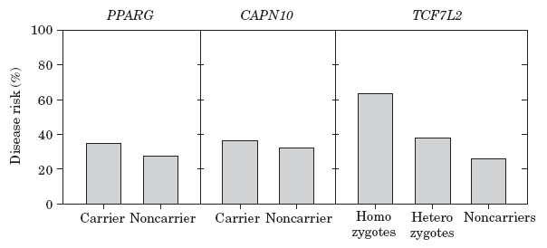
[A text description of this chart is also available.]
Figure 22-3
Disease risks associated with profiles in genetic testing for multifactorial disorders (10). Bars indicate the frequency distribution of the genetic profiles quantified by the number of risk genotypes in the profile. The frequencies are presented on the left axis. The scatter plots represent the disease risks associated with the genetic profiles when all individual variants have the same odds ratio (OR = 1.5; Figure 22.3b) and when the odds ratios vary (Figure 22.3c). The disease risks are presented on the right axis. Genetic profiles were constructed using a previously described method (15). We assumed that the disease risk in the population was 10% and that the frequencies of the risk genotypes varied between 1% and 60% (incremental from 1% to 19% by 1% and from 20% to 60% by 2%). In Figure 22.3c we assumed that the odds ratios varied from 1.05 to 2.0 (incremental from 1.05 to 1.19 by 0.01 and from 1.20 to 2.0 by 0.05).
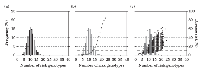
[A text description of this chart is also available.]
- Page last reviewed: January 6, 2010 (archived document)
- Content source: