

MERS-CoV Photos
 ShareCompartir
Coronaviruses derive their name from the fact that under electron microscopic examination, each virion is surrounded by a “corona,” or halo. This is due to the presence of viral spike peplomers emanating from each proteinaceous envelope.
Click on image to enlarge.
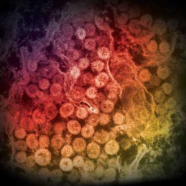
Image source: Cynthia Goldsmith/Azaibi Tamin
An electron micrograph of a thin section of MERS-CoV, showing the spherical particles within the cytoplasm of an infected cell.
Click on image to enlarge.
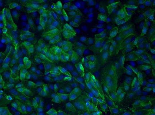
Image source: Jennifer L. Harcourt
Human serum antibodies react with MERS-CoV-infected Vero cells, indicating the patient has been infected with MERS-CoV.
Click on image to enlarge.
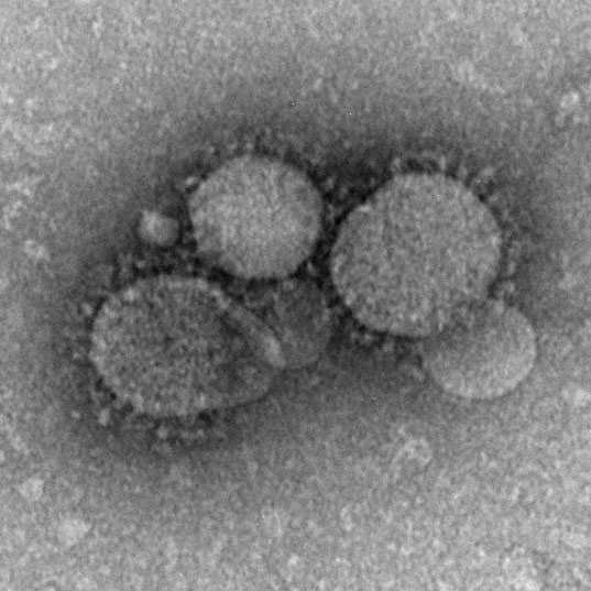
Image source: Cynthia Goldsmith/Maureen Metcalfe/Azaibi Tamin
MERS-CoV particles as seen by negative stain electron microscopy. Virions contain characteristic club-like projections emanating from the viral membrane.
Click on image to enlarge.
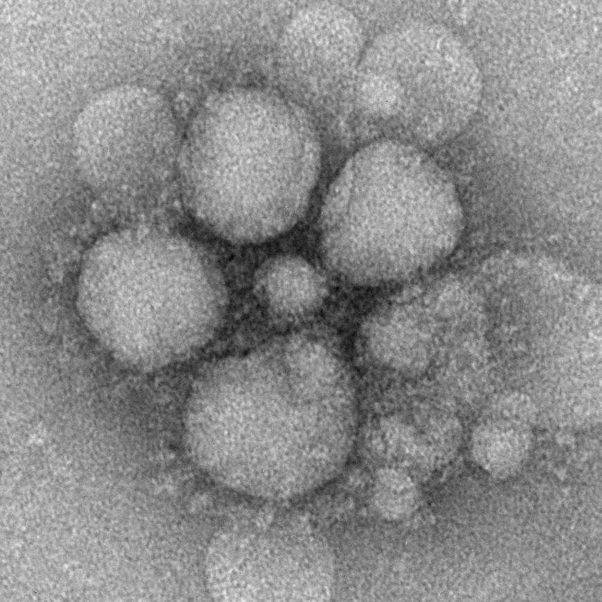
Image source: Cynthia Goldsmith/Maureen Metcalfe/Azaibi Tamin
MERS-CoV particles as seen by negative stain electron microscopy. Virions contain characteristic club-like projections emanating from the viral membrane.
Click on image to enlarge.
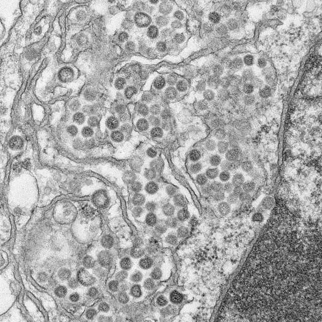
Image source: Maureen Metcalfe/Azaibi Tamin
An electron micrograph of a thin section of MERS-CoV, showing the spherical particles and cross-sections through the viral nucleocapsid.
Click on image to enlarge.
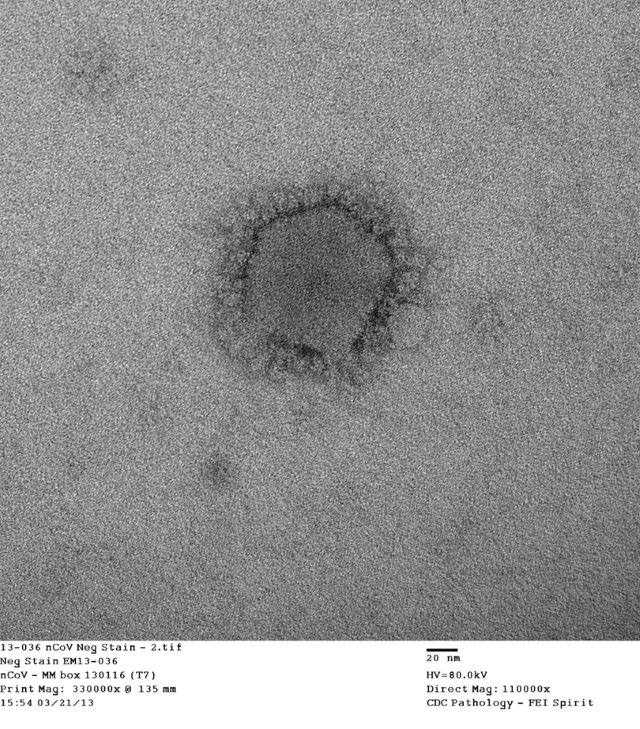
Image source: Cynthia Goldsmith/Azaibi Tamin
Negative stain electron microscopy shows a MERS-CoV particle with club-shaped surface projections surrounding the periphery of the particle, a characteristic feature of coronaviruses.
- Page last reviewed: September 14, 2017
- Page last updated: May 28, 2014
- Content source: