Smegma
Smegma (Greek smēgma[1]) is a combination of shed skin cells, skin oils, and moisture. It occurs in both male and female mammalian genitalia. In female bodies, it collects around the clitoris and in the folds of the labia minora; in males, smegma collects under the foreskin.
Females

The accumulation of sebum combined with dead skin cells forms smegma. Smegma clitoridis is defined as the secretion of the apocrine glands of the clitoris, in combination with desquamating epithelial cells.[2] Glands that are located around the clitoris and the vulva majoris secrete sebum. If smegma is not removed frequently it can lead to clitoral adhesion which can make clitoral stimulation (such as masturbation) painful (clitorodynia).
Males
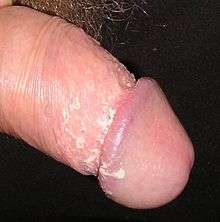
In males, smegma helps keep the glans moist and facilitates sexual intercourse by acting as a lubricant.[3][4][5] Smegma is completely benign.[6]
Smegma was originally thought to be produced by sebaceous glands near the frenulum called Tyson's glands; however, subsequent studies have failed to find these glands.[7] Wright states that smegma is produced from minute microscopic protrusions of the mucosal surface of the foreskin and that living cells constantly grow towards the surface, undergo fatty degeneration, separate off, and form smegma.[3] Parkash et al. found that smegma contains 26.6% fats and 13.3% proteins, which they judged to be consistent with necrotic epithelial debris.[7]
Newly produced smegma has a smooth, moist texture. It is thought to be rich in squalene[8] and contain prostatic and seminal secretions, desquamated epithelial cells, and the mucin content of the urethral glands of Littré.[5] Smegma contains cathepsin B, lysozymes,[9] chymotrypsin, neutrophil elastase and cytokines, which aid the immune system.[10]
According to Wright, little smegma is produced during childhood, although the foreskin may contain sebaceous glands. She also says that production of smegma increases from adolescence until sexual maturity when the function of smegma for lubrication assumes its full value, and from middle-age production starts to decline and in old age virtually no smegma is produced.[3] Øster reported that the incidence of smegma increased from 1% among 6- to 7-year-olds and 8- to 9-year-olds to 8% among 14- to 15-year-olds and 16- to 17-year-olds (an overall incidence of 5%).[11]
There is no evidence that smegma causes penile cancer,[4] but its presence over a long period of time may irritate and inflame the penis,[3] which may increase the risk of cancer. It may also make it harder to see very early cancers.[12]
Other animals
In healthy animals, smegma helps clean and lubricate the genitals. In veterinary medicine, analysis of this smegma is sometimes used for detection of urogenital tract pathogens, such as Tritrichomonas foetus.[13] Accumulation of smegma in the equine preputial folds and the urethral fossa and urethral diverticulum can form large "beans" and promote the carriage of Taylorella equigenitalis, the causative agent of contagious equine metritis.[14] Some equine veterinarians have recommended periodic cleaning of male genitals to improve the health of the animal.[15]
See also
- List of cutaneous conditions
- Mycobacterium smegmatis – found in smegma
- Keratin pearl
Notes
- "smegma". The Merriam-Webster.com Dictionary. Merriam-Webster, Inc. Retrieved 2017-03-02.
- "Medical Dictionary". Medilexicon.
- Wright J (September 1970). "How smegma serves the penis: Nature's assurance that the uncircumcised glans penis will function smoothly is provided by smegma". Sexology. 37 (2): 50–53.
- Van Howe RS, Hodges FM (October 2006). "The carcinogenicity of smegma: debunking a myth". Journal of the European Academy of Dermatology and Venereology. 20 (9): 1046–1054. doi:10.1111/j.1468-3083.2006.01653.x. PMID 16987256.
- Fleiss PM, Hodges FM, Van Howe RS (October 1998). "Immunological functions of the human prepuce". Sexually Transmitted Infections. 74 (5): 364–367. doi:10.1136/sti.74.5.364. PMC 1758142. PMID 10195034.
- McGregor TB, Pike JG, Leonard MP (2007). "Pathologic and physiologic phimosis: Approach to the phimotic foreskin". Canadian Family Physician. 53 (3): 445–8. PMC 1949079. PMID 17872680.
- Parkash S, Jeyakumar S, Subramanyan K, Chaudhuri S (August 1973). "Human subpreputial collection: its nature and formation". Journal of Urology. 110 (2): 211–212. doi:10.1016/s0022-5347(17)60164-2. PMID 4722614.
- O'Neill HJ, Gershbein LL (1976). "Lipids of human and equine smegma". Oncology. 33 (4): 161–166. doi:10.1159/000225134. PMID 1018879.
- Frohlich E, Schaumburg-Lever G, Klessen C (1993). "Immunelectron microscopic localization of cathepsin B in human exocrine glands". Journal of Cutaneous Pathology. 20 (1): 54–60. doi:10.1111/j.1600-0560.1993.tb01250.x. PMID 8468418.
- Chukwuemeka Anyanwu L, Kashibu E, Edwin CP, Mohammad AM (2012). "Microbiology of smegma in boys in Kano, Nigeria". Journal of Surgical Research. 173 (1): 21–25. doi:10.1016/j.jss.2011.04.057. PMID 21872267.
- Oster J (April 1968). "Further fate of the foreskin. Incidence of preputial adhesions, phimosis, and smegma among Danish schoolboys". Archives of Disease in Childhood. 43 (228): 200–3. doi:10.1136/adc.43.228.200. PMC 2019851. PMID 5689532.
- "What are the risk factors for penile cancer?". American Cancer Society. Retrieved 14 February 2014.
- Chen XG, Li J (2001). "Increasing the sensitivity of PCR detection in bovine preputial smegma spiked with Tritrichomonas foetus by the addition of agar and resin". Parasitology Research. 87 (7): 556–558. doi:10.1007/s004360100401. PMID 11484853.
- Primary Industries Ministerial Council of Australia and New Zealand (2002). Disease strategy: Contagious equine metritis (Version 1.0). In: Australian Veterinary Emergency Plan (AUSVETPLAN), Edition 3, PIMCANZ, Canberra, ACT.
- Michael Lowder (September 1, 2001). "A Clean Sheath Is A Healthy Sheath Archived 2005-09-14 at the Wayback Machine". Horse City. Retrieved on September 4, 2008.
External links
