Vernix caseosa
Vernix caseosa, also known as vernix, is the waxy or cheese-like white substance found coating the skin of newborn human babies. It is produced by dedicated cells and is thought to have some protective roles during fetal development and for a few hours after birth.
Etymology
In Latin, vernix means varnish and caseosa means cheesy.
Characteristics
Composition
Vernix has a highly variable makeup but is primarily composed of sebum, cells that have sloughed off the fetus's skin and shed lanugo hair.[1] 12% of the dry weight of vernix is composed of branched chain fatty acids,[2] cholesterol, and ceramide. Vernix of term infants has more squalene and a higher wax ester to sterol ester ratio than preterm infants.[1]
Comparison of lipid components of vernix caseosa, stratum corneum and skin surface (sebaceous):[3][4]
| Lipid fractions | Vernix caseosa lipids | Stratum corneum lipids | Skin surface lipids |
|---|---|---|---|
| Cholesterol esters | 30.6 | - | 3.0 |
| Ceramides | 17.9 | 40.0 | - |
| Triglycerides | 15.1 | - | 41.8 |
| Cholesterol | 7.5 | 25.0 | - |
| Free fatty acids | 6.5 | 25.0 | 18.4 |
| Phospholipids | 6.1 | - | 1.5 |
| Wax esters | 6.0 | - | 20.3 |
| Squalene | 4.0 | - | 12.2 |
| Wax diesters | 3.7 | - | - |
| Cerebrosides | 2.4 | - | - |
| Cholesterol sulfate | 0.3 | 10.0 | - |
| Alkanes | - | - | 2.8 |
Amino acid composition of vernix caseosa:[4][5]
| Amino acid | Percent |
|---|---|
| Asparagine | 34.7 |
| Glutamine | 22.7 |
| Proline | 14.9 |
| Cysteine | 7.9 |
| Alanine | 7.4 |
| Leucine | 5.3 |
| Valine | 3.7 |
| Methionine | 3.4 |
Morphology
Cells of vernix are typically polygonal or ovoid in shape and lack nuclei. Nuclear ghosts are frequently observed. Vernix corneocytes lack desmosomal attachment and this distinguishes them from corneocytes found in mature stratum corneum.[6] Thickness of a corneocyte is 1-2 µm. These cells are surrounded by a layer of amorphous lipids lacking typical lamellar architecture present in mature stratum corneum.[4]
Physical properties
Vernix is not uniformly distributed, but rather present in form of cellular sponges. The critical surface tension of vernix is 39 dyne/cm.[7] Despite its water content (82%), vernix is nonpolar. These features point towards the "waterproofing" function of vernix, thereby preventing heat loss soon after birth.[4]
Biological properties
Vernix provides electrical isolation for the fetus,[8] which is presumably an important aspect of developing fetal anatomy.[4] Early scientific studies indicated increased evaporative heat loss in infants when vernix was removed soon after birth;[9] but newer reports confirm that washing skin surface after birth reduces evaporative water losses compared to surface of newborns in which vernix is left in situ.[10] Vernix is hydrophobic. Vernix is believed to assist in the development of the human intestinal microbiota.[2]
Secretion
The sebum in vernix is produced in utero by the sebaceous glands around the 20th week of gestation. Vernix appears primarily in full term infants, while premature and postmature births generally do not display any.[1] Postdates desquamation (flakey skin in babies born >42 weeks) is thought to be due to loss of vernix.
Functions
Vernix is theorized to serve several purposes, including moisturizing the infant's skin, and facilitating passage through the birth canal. It serves to conserve heat and protect the delicate newborn skin from environmental stress. Vernix is also thought to have an antibacterial effect;[4] though there is little evidence to support a chemical role of vernix in protecting the infant from infection, it may form a physical barrier to the passage of bacteria.[1]
Non-human observations
In 2018, Tom Brenna at Cornell University published an account of vernix-like material obtained (with the help of San Diego Seaworld) from pups of the California sea lion (Zalophus californianus).[11] Mass spectrometry of the material showed it to be fundamentally the same as human vernix, in both BCFA (branch-chain fatty acids) and squalene content. In their study, the presence of vernix throughout the infant gastro-intestinal tract, as well as in the meconium (first excretion), in both human and sea lion neonates, argues that the function of vernix may not be as an external skin protection, as often described in the literature, but as a preparation of the newborn GI tract against water-borne bacteria. A potential cause of fatality in premature human infants is necrotizing enterocolitis, which occurs when the foetal ingestion of its own vernix along with the amniotic fluid has not been completed.
Additional images
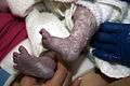 Vernix on a newborn's legs and feet.
 Traces of vernix caseosa on a full term newborn.
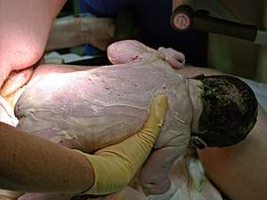
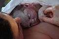 Closeup of baby's face right after birth, skin covered in vernix and some blood.
References
- Schachner, Lawrence A.; Hansen, Ronald C. (2003). Pediatric dermatology. St. Louis: Mosby. pp. 206–7. ISBN 978-0-323-02611-6.
- Ran-Ressler RR, Devapatla S, Lawrence P, Brenna JT (2008). "Branched chain fatty acids are constituents of the normal healthy newborn gastrointestinal tract" (PDF). Pediatric Research. 64 (6): 605–609. doi:10.1203/PDR.0b013e318184d2e6. PMC 2662770. PMID 18614964.CS1 maint: multiple names: authors list (link)
- Sumida Y, Yakumaru M, Tokitsu Y, et al. Studies on the function of Vernix caseosa: The secrecy of Baby's skin. Cannes, France: International Federation of the Societies of Cosmetic Chemists 20th International Conference; 1998. pp. 1–7.
- Hoath, Steven (2003). Neonatal skin : structure and function (2. ed., rev. and expanded. ed.). New York [u.a.]: Dekker. pp. 193–208. ISBN 0-8247-0887-3.
- Baker, SM; Balo, NN; Abdel Aziz, FT (Mar–Apr 1995). "Is vernix caseosa a protective material to the newborn? A biochemical approach". Indian Journal of Pediatrics. 62 (2): 237–9. doi:10.1007/bf02752334. PMID 10829874.
- Pickens, WL; Warner, RR; Boissy, YL; Boissy, RE; Hoath, SB (Nov 2000). "Characterization of vernix caseosa: water content, morphology, and elemental analysis". The Journal of Investigative Dermatology. 115 (5): 875–81. doi:10.1046/j.1523-1747.2000.00134.x. PMID 11069626.
- Youssef, W; Wickett, RR; Hoath, SB (Feb 2001). "Surface free energy characterization of vernix caseosa. Potential role in waterproofing the newborn infant". Skin Research and Technology. 7 (1): 10–7. doi:10.1034/j.1600-0846.2001.007001010.x. PMID 11301635.
- Wakai, RT; Lengle, JM; Leuthold, AC (Jul 2000). "Transmission of electric and magnetic foetal cardiac signals in a case of ectopia cordis: the dominant role of the vernix. caseosa". Physics in Medicine and Biology. 45 (7): 1989–95. doi:10.1088/0031-9155/45/7/320. PMID 10943933.
- Saunders, Colman (1 August 1948). "The vernix caseosa and subnormal temperature in premature infants". The Journal of Obstetrics and Gynaecology of the British Empire. 55 (4): 442–444. doi:10.1111/j.1471-0528.1948.tb07409.x. PMID 18878967.
- Riesenfeld B, Stromberg B, Sedin G. The influence of vernix caseosa on water transport through semipermeable membranes and the skin of full-term infants. Neonatal Physiological Measurements: Proceedings of the Second International Conference on Fetal and Neonatal Physiological Measurements, 1984:3–6.
- Brenna, Tom (May 10, 2018). "Sea Lions Develop Human-like Vernix Caseosa Delivering Branched Fats and Squalene to the GI Tract". Scientific Reports. 8 (7478): 7478. doi:10.1038/s41598-018-25871-1. PMC 5945841. PMID 29748625.
Further reading
| Wikimedia Commons has media related to Vernix caseosa. |
- Sarkar, Rashmi; Basu, Srikanta; Agrawal, R K; Gupta, Piyush (2010). "Skin Care for the Newborn" (PDF). Indian Pediatrics. 47 (7): 593–8. doi:10.1007/s13312-010-0132-0. PMID 20683112.
- Visscher, Marty O; Narendran, Vivek; Pickens, William L; Laruffa, Angela A; Meinzen-Derr, Jareen; Allen, Kathleen; Hoath, Steven B (2005). "Vernix Caseosa in Neonatal Adaptation". Journal of Perinatology. 25 (7): 440–6. doi:10.1038/sj.jp.7211305. PMID 15830002.
- Haubrich, Kathleen A. (2003). "Role of Vernix Caseosa in the Neonate: Potential Application in the Adult Population". AACN Advanced Critical Care. 14 (4): 457–64. doi:10.1097/00044067-200311000-00006. PMID 14595204.