NUT midline carcinoma
NUT carcinoma (formerly NUT midline carcinoma), is a rare genetically defined, very aggressive squamous cell epithelial cancer that usually arises in the midline of the body and is characterized by a chromosomal rearrangement in the nuclear protein in testis gene.[2] In approximately 75% of cases, the coding sequence of NUTM1 on chromosome 15q14 is fused to BRD4 or BRD3, which creates a chimeric gene that encodes the BRD-NUT fusion protein. The remaining cases, the fusion of NUTM1 is to an unknown partner gene, usually called NUT-variant.
| NUT midline carcinoma | |
|---|---|
| Other names | NMC[1] |
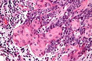 | |
| Micrograph of a NUT midline carcinoma with the characteristic well-differentiated islands of squamous epithelium. H&E stain. | |
| Specialty | Oncology |
Signs and symptoms
In the United States, about 20-30 cases are reported each year. This may be a gross underestimate of the total number of cases as few laboratories have the reagents and expertise to make the diagnosis.[3] The symptoms are similar to other forms of cancer and dependent on the stage. While generalized symptoms (weight loss and fatigue) may be seen, site specific symptoms are also present. If the tumor involves the head and neck region (in about 35%), then pain, a mass, obstructive symptoms, among others, may be experienced. NUT midline carcinomas are not specific to any tissue type or organ.[4] Common sites include the head, neck and mediastinum.[5] The median age at diagnosis is 17 years, but older patients may be affected.
Diagnosis
NMC when viewed microscopically, are poorly differentiated carcinomas which show abrupt transitions to islands of well-differentiated squamous epithelium.[4][5] This tumor pattern is not specific or unique to NUT midline carcinoma, but this pattern is most suggestive of the diagnosis. The neoplastic cells will show a positive reaction with various cytokeratins, p63, CEA, and CD34 immunohistochemistry. However, the NUT antibody confirms the diagnosis (although only available in a limited number of laboratories).
Differential diagnosis
The differential diagnosis is quite wide, but it is important to consider this tumor type when seeing a poorly differentiated tumor that shows abrupt areas of keratinization. Other tumors included in the differential diagnosis are sinonasal undifferentiated carcinomas, Ewing sarcoma/Primitive neuroectodermal tumor, leukemia, rhabdomyosarcoma, and melanoma. When NUT midline carcinoma is seen in the head and neck, the squamous lining of the cavities may be entrapped by the neoplastic cells, and so it is important to document the carcinoma cells in the rest of the tumor by a variety of stains (including cytokeratin or p63). One of the most helpful and characteristic findings is the focal abrupt squamous differentiation, where stratification and gradual differentiation are absent, resembling a Hassall corpuscle of the thymus.[6]
The defining feature of NMCs is rearrangement of the NUT gene.[4] Most common is a translocation involving the BRD4 gene and NUT gene (t(15;19)(q13;p13.1)).[5][7]
Treatment
BET inhibitors are used for treatment
Prognosis
NUT midline carcinoma is very resistant to standard chemotherapy treatments. The tumor may initially respond to therapy, and then rapid recurrence is experienced. A multimodality approach to treatment is advocated, especially since most patients present with advanced disease. Treatment must be tailored to the individual patient, with several promising new targeted molecular therapies in clinical trials. Specific molecular targeted therapies (BET inhibitors and histone deacetylase inhibitors (HDACi)) may help to yield growth arrest of the neoplastic cells.[6] Overall, there is a mean survival of 6–9 months.[8][9]
See also
- BET inhibitor
- Mediastinum
References
- RESERVED, INSERM US14-- ALL RIGHTS. "Orphanet: NUT midline carcinoma". www.orpha.net. Retrieved 18 November 2019.
- Online Mendelian Inheritance in Man (OMIM) 608963
- "International NUT Midline Carcinoma Registry - Doctors".
- French, CA. (Nov 2010). "NUT midline carcinoma". Cancer Genet Cytogenet. 203 (1): 16–20. doi:10.1016/j.cancergencyto.2010.06.007. PMC 3000636. PMID 20951314.
- French, CA. (Jun 2010). "Demystified molecular pathology of NUT midline carcinomas". J Clin Pathol. 63 (6): 492–6. doi:10.1136/jcp.2007.052902. PMID 18552174.
- French, C. A. (2013). "The importance of diagnosing NUT midline carcinoma". Head and Neck Pathology. 7 (1): 11–6. doi:10.1007/s12105-013-0428-1. PMC 3597165. PMID 23463074.
- Online Mendelian Inheritance in Man (OMIM) 608749
- NEJM 367:647 doi:10.1056/NEJMra1112635
- "International NUT Midline Carcinoma Registry - Doctors".