Chromosomal translocation
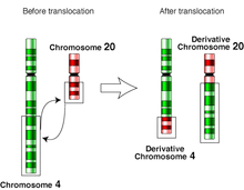
In genetics, chromosome translocation is a phenomenon that results in unusual rearrangement of chromosomes. This includes balanced and unbalanced translocation, with two main types: reciprocal-, and Robertsonian translocation. Reciprocal translocation is a chromosome abnormality caused by exchange of parts between non-homologous chromosomes. Two detached fragments of two different chromosomes are switched. Robertsonian translocation occurs when two non-homologous chromosomes get attached, meaning that given two healthy pairs of chromosomes, one of each pair "sticks" together.[1]

A gene fusion may be created when the translocation joins two otherwise-separated genes. It is detected on cytogenetics or a karyotype of affected cells. Translocations can be balanced (in an even exchange of material with no genetic information extra or missing, and ideally full functionality) or unbalanced (where the exchange of chromosome material is unequal resulting in extra or missing genes).[2][3]
Reciprocal translocations
Reciprocal translocations are usually an exchange of material between non-homologous chromosomes. Estimates of incidence range from about 1 in 500[4] to 1 in 625 human newborns.[5] Such translocations are usually harmless and may be found through prenatal diagnosis. However, carriers of balanced reciprocal translocations have increased risks of creating gametes with unbalanced chromosome translocations, leading to Infertility, miscarriages or children with abnormalities. Genetic counseling and genetic testing are often offered to families that may carry a translocation. Most balanced translocation carriers are healthy and do not have any symptoms.
It is important to distinguish between chromosomal translocations occurring in gametogenesis, due to errors in meiosis, and translocations that occur in cellular division of somatic cells, due to errors in mitosis. The former results in a chromosomal abnormality featured in all cells of the offspring, as in translocation carriers. Somatic translocations, on the other hand, result in abnormalities featured only in the affected cell line, as in chronic myelogenous leukemia with the Philadelphia chromosome translocation.
Nonreciprocal translocation
Nonreciprocal translocation involves the one-way transfer of genes from one chromosome to another nonhomologous chromosome.[6]
Robertsonian translocations
Robertsonian translocation is a type of translocation caused by breaks at or near the centromeres of two acrocentric chromosomes. The reciprocal exchange of parts gives rise to one large metacentric chromosome and one extremely small chromosome that may be lost from the organism with little effect because it contains few genes. The resulting karyotype in humans leaves only 45 chromosomes, since two chromosomes have fused together.[7] This has no direct effect on the phenotype, since the only genes on the short arms of acrocentrics are common to all of them and are present in variable copy number (nucleolar organiser genes).
Robertsonian translocations have been seen involving all combinations of acrocentric chromosomes. The most common translocation in humans involves chromosomes 13 and 14 and is seen in about 0.97 / 1000 newborns.[8] Carriers of Robertsonian translocations are not associated with any phenotypic abnormalities, but there is a risk of unbalanced gametes that lead to miscarriages or abnormal offspring. For example, carriers of Robertsonian translocations involving chromosome 21 have a higher risk of having a child with Down syndrome. This is known as a 'translocation Downs'. This is due to a mis-segregation (nondisjunction) during gametogenesis. The mother has a higher (10%) risk of transmission than the father (1%). Robertsonian translocations involving chromosome 14 also carry a slight risk of uniparental disomy 14 due to trisomy rescue.
Role in disease
Some human diseases caused by translocations are:
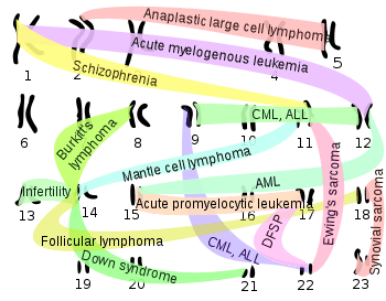
- Cancer: Several forms of cancer are caused by acquired translocations (as opposed to those present from conception); this has been described mainly in leukemia (acute myelogenous leukemia and chronic myelogenous leukemia). Translocations have also been described in solid malignancies such as Ewing's sarcoma.
- Infertility: One of the would-be parents carries a balanced translocation, where the parent is asymptomatic but conceived fetuses are not viable.
- Down syndrome is caused in a minority (5% or less) of cases by a Robertsonian translocation of the chromosome 21 long arm onto the long arm of chromosome 14.[9]
Chromosomal translocations between the sex chromosomes can also result in a number of genetic conditions, such as
- XX male syndrome: caused by a translocation of the SRY gene from the Y to the X chromosome
By chromosome

ALL – Acute lymphoblastic leukemia
AML – Acute myeloid leukemia
CML – Chronic myelogenous leukemia
DFSP – Dermatofibrosarcoma protuberans
Denotation
The International System for Human Cytogenetic Nomenclature (ISCN) is used to denote a translocation between chromosomes.[11] The designation t(A;B)(p1;q2) is used to denote a translocation between chromosome A and chromosome B. The information in the second set of parentheses, when given, gives the precise location within the chromosome for chromosomes A and B respectively—with p indicating the short arm of the chromosome, q indicating the long arm, and the numbers after p or q refers to regions, bands and subbands seen when staining the chromosome with a staining dye.[12] See also the definition of a genetic locus. The translocation is the mechanism that can cause a gene to move from one linkage group to another.
Examples
| Translocation | Associated diseases | Fused genes/proteins | |
|---|---|---|---|
| First | Second | ||
| t(8;14)(q24;q32) | Burkitt's lymphoma | c-myc on chromosome 8, gives the fusion protein lymphocyte-proliferative ability | IGH@ (immunoglobulin heavy locus) on chromosome 14, induces massive transcription of fusion protein |
| t(11;14)(q13;q32) | Mantle cell lymphoma[13] | cyclin D1[13] on chromosome 11, gives fusion protein cell-proliferative ability | IGH@[13] (immunoglobulin heavy locus) on chromosome 14, induces massive transcription of fusion protein |
| t(14;18)(q32;q21) | Follicular lymphoma (~90% of cases)[14] | IGH@[13] (immunoglobulin heavy locus) on chromosome 14, induces massive transcription of fusion protein | Bcl-2 on chromosome 18, gives fusion protein anti-apoptotic abilities |
| t(10;(various))(q11;(various)) | Papillary thyroid cancer[15] | RET proto-oncogene[15] on chromosome 10 | PTC (Papillary Thyroid Cancer) – Placeholder for any of several other genes/proteins[15] |
| t(2;3)(q13;p25) | Follicular thyroid cancer[15] | PAX8 – paired box gene 8[15] on chromosome 2 | PPARγ1[15] (peroxisome proliferator-activated receptor γ 1) on chromosome 3 |
| t(8;21)(q22;q22)[14] | Acute myeloblastic leukemia with maturation | ETO on chromosome 8 | AML1 on chromosome 21 found in ~7% of new cases of AML, carries a favorable prognosis and predicts good response to cytosine arabinoside therapy[14] |
| t(9;22)(q34;q11) Philadelphia chromosome | Chronic myelogenous leukemia (CML), acute lymphoblastic leukemia (ALL) | Abl1 gene on chromosome 9[16] | BCR ("breakpoint cluster region" on chromosome 22[16] |
| t(15;17)(q22;q21)[14] | Acute promyelocytic leukemia | PML protein on chromosome 15 | RAR-α on chromosome 17 persistent laboratory detection of the PML-RARA transcript is strong predictor of relapse[14] |
| t(12;15)(p13;q25) | Acute myeloid leukemia, congenital fibrosarcoma, secretory breast carcinoma, mammary analogue secretory carcinoma of salivary glands, cellular variant of mesoblastic nephroma | TEL on chromosome 12 | TrkC receptor on chromosome 15 |
| t(9;12)(p24;p13) | CML, ALL | JAK on chromosome 9 | TEL on chromosome 12 |
| t(12;16)(q13;p11) | Myxoid liposarcoma | DDIT3 (formerly CHOP) on chromosome 12 | FUS gene on chromosome 16 |
| t(12;21)(p12;q22) | ALL | TEL on chromosome 12 | AML1 on chromosome 21 |
| t(11;18)(q21;q21) | MALT lymphoma[17] | BIRC3 (API-2) | MLT[17] |
| t(1;11)(q42.1;q14.3) | Schizophrenia[10] | ||
| t(2;5)(p23;q35) | Anaplastic large cell lymphoma | ALK | NPM1 |
| t(11;22)(q24;q11.2-12) | Ewing's sarcoma | FLI1 | EWS |
| t(17;22) | DFSP | Collagen I on chromosome 17 | Platelet derived growth factor B on chromosome 22 |
| t(1;12)(q21;p13) | Acute myelogenous leukemia | ||
| t(X;18)(p11.2;q11.2) | Synovial sarcoma | ||
| t(1;19)(q10;p10) | Oligodendroglioma and oligoastrocytoma | ||
| t(17;19)(q22;p13) | ALL | ||
| t(7,16) (q32-34;p11) or t(11,16) (p11;p11) | Low-grade fibromyxoid sarcoma | FUS | CREB3L2 or CREB3L1 |
History
In 1938, Karl Sax, at the Harvard University Biological Laboratories, published a paper entitled "Chromosome Aberrations Induced by X-rays", which demonstrated that radiation could induce major genetic changes by affecting chromosomal translocations. The paper is thought to mark the beginning of the field of radiation cytology, and led him to be called "the father of radiation cytology".
See also
- Accipitridae
- Aneuploidy
- Chromosome abnormalities
- DbCRID
- Fusion gene
- Takifugu rubripes
References
- "EuroGentest: chromosome_translocations". www.eurogentest.org. Retrieved March 29, 2019.
- "EuroGenTest Chromosome Translocations". Retrieved March 5, 2016.
- "Balanced translocation". Retrieved March 4, 2016.
- Caroline Mackie Ogilvie; Paul N Scriven (December 2002). "Meiotic outcomes in reciprocal translocation carriers ascertained in 3-day human embryos". European Journal of Human Genetics. 10 (12): 801–806. doi:10.1038/sj.ejhg.5200895. PMID 12461686.
- M. Oliver-Bonet; J. Navarro1; M. Carrera; J. Egozcue; J. Benet (October 2002). "Aneuploid and unbalanced sperm in two translocation carriers: evaluation of the genetic risk". Molecular Human Reproduction. 8 (10): 958–963. doi:10.1093/molehr/8.10.958. ISSN 1460-2407. PMID 12356948. Retrieved December 26, 2008.
- "Translocation". Carmel Clay Schools. Archived from the original on December 1, 2017. Retrieved March 2, 2009.
- Hartwell, Leland H. (2011). Genetics: From Genes to Genomes. New York: McGraw-Hill. p. 443. ISBN 978-0-07-352526-6.
- E. Anton; J. Blanco; J. Egozcue; F. Vidal (April 29, 2004). "Sperm FISH studies in seven male carriers of Robertsonian translocation t(13;14)(q10;q10)". Human Reproduction. 19 (6): 1345–1351. doi:10.1093/humrep/deh232. ISSN 1460-2350. PMID 15117905. Retrieved December 25, 2008.
- "Causes". nhs.uk. Retrieved March 16, 2018.
- Semple CA, Devon RS, Le Hellard S, Porteous DJ (April 2001). "Identification of genes from a schizophrenia-linked translocation breakpoint region". Genomics. 73 (1): 123–6. doi:10.1006/geno.2001.6516. PMID 11352574.
- Schaffer, Lisa. (2005) International System for Human Cytogenetic Nomenclature S. Karger AG ISBN 978-3-8055-8019-9
- "Characteristics of chromosome groups: Karyotyping". rerf.jp. Radiation Effects Research Foundation. Retrieved June 30, 2014.
- Li JY, Gaillard F, Moreau A, et al. (May 1999). "Detection of translocation t(11;14)(q13;q32) in mantle cell lymphoma by fluorescence in situ hybridization". Am. J. Pathol. 154 (5): 1449–52. doi:10.1016/S0002-9440(10)65399-0. PMC 1866594. PMID 10329598.
- Burtis, Carl A.; Ashwood, Edward R.; Bruns, David E. (December 16, 2011). "44. Hematopoeitic malignancies". Tietz Textbook of Clinical Chemistry and Molecular Diagnostics. Elsevier Health Sciences. pp. 1371–1396. ISBN 978-1-4557-5942-2. Retrieved November 5, 2012.
- Chapter 20 in: Mitchell, Richard Sheppard; Kumar, Vinay; Abbas, Abul K.; Fausto, Nelson (2007). Robbins Basic Pathology. Philadelphia: Saunders. ISBN 978-1-4160-2973-1. 8th edition.
- Kurzrock R, Kantarjian HM, Druker BJ, Talpaz M (May 2003). "Philadelphia chromosome-positive leukemias: from basic mechanisms to molecular therapeutics". Ann. Intern. Med. 138 (10): 819–30. doi:10.7326/0003-4819-138-10-200305200-00010. PMID 12755554.
- Page 626 in: Mitchell, Richard Sheppard; Kumar, Vinay; Abbas, Abul K.; Fausto, Nelson (2007). Robbins Basic Pathology. Philadelphia: Saunders. ISBN 978-1-4160-2973-1. 8th edition.
| Wikimedia Commons has media related to Chromosomal translocations. |