LE cell
An LE cell (Lupus Erythematosus cell) is a neutrophil or macrophage that has phagocytized (engulfed) the denatured nuclear material of another cell.[1] The denatured material is an absorbed hematoxylin body (also called an LE body).[2]
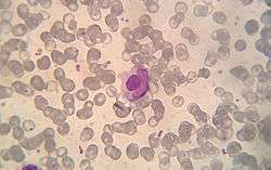
They are a characteristic of lupus erythematosus,[3] but also found in similar connective tissue disorders or some autoimmune diseases like in severe rheumatoid arthritis. LE cells can be observed in drug-induced lupus, for example, following treatment with methyldopa.[4]
The LE cell was discovered in bone marrow in 1948 by Malcolm McCallum Hargraves (1903–1982), a Physician and Practicing Histologist at the Mayo Clinic.[5] Hargraves may have gained priority by suppressing a publication draft of John R. Haserick, who credits Dr. Dorothy Sundberg, chief hematologist at the University of Minnesota Hospitals, with first identifying LE cells. [6]
Classically, the LE cell is analyzed microscopically, but it is also possible to investigate this phenomenon by flow cytometry.[7]
LE cells shouldn't be confused with Tart cells which have engulfed nuclear material, but with a visible lchromatin rather than homogenous appearance. [8]
References
- "Medical Definition of LE CELL". www.merriam-webster.com.
- similima.com > Autoimmunity Archived 2011-07-27 at the Wayback Machine By Muhammed Muneer. Retrieved Mars 2011
- "Archived copy". Archived from the original on 2012-03-08. Retrieved 2010-06-28.CS1 maint: archived copy as title (link)
- Cheesbrough, Monica (2000-10-26). District Laboratory Practice in Tropical Countries. Cambridge University Press. ISBN 9780521665452.
- Hargraves M, Richmond H, Morton R. Presentation of two bone marrow components, the tart cell and the LE cell. Mayo Clin Proc 1948;27:25–28.
- https://www.derm101.com/dpc-archive/april-june-2000-volume-6-no-2/dpc0602a20-discovery-of-the-le-factor/
- Böhm, Ingrid (1 January 2004). "Flow Cytometric Analysis of the LE Cell Phenomenon". Autoimmunity. 37 (1): 37–44. doi:10.1080/08916930310001630325. PMID 15115310.
- Li, Qing Kay; Khalbuss, Walid E. (2015). Diagnostic Cytopathology Board Review and Self-Assessment. Springer, New York, NY. p. 179. doi:10.1007/978-1-4939-1477-7_2. ISBN 9781493914760.