Hip prosthesis zones
After hip replacement, hip prosthesis zones are regions in the interface between prosthesis material and the surrounding bone. These are used as reference regions when describing for example complications including hip prosthesis loosening on medical imaging. Postoperative controls after hip replacement surgery is routinely done by projectional radiography in anteroposterior and lateral views.
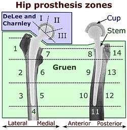
DeLee and Charnley
The DeLee and Charnley system applies to the acetabular cup on anteroposterior radiographs. It divides the acetabulum into three equally large zones.[1]
Gruen
The Gruen zones is a system of dividing the interface between the bone and the stem of the hip prosthesis.
| Zone | Description |
|---|---|
| I | Greater trochanter |
| II | |
| III | |
| IV | Distal to tip |
| V | |
| VI | |
| VII | Lesser trochanter |
References
- Page 958 in John J. Callaghan, Aaron G. Rosenberg, Harry E. Rubash (2007). The Adult Hip, Volume 1. Lippincott Williams & Wilkins. ISBN 9780781750929.CS1 maint: multiple names: authors list (link)
- Neumann, Daniel R.P.; Thaler, Christoph; Hitzl, Wolfgang; Huber, Monika; Hofstädter, Thomas; Dorn, Ulrich (2010). "Long-Term Results of a Contemporary Metal-on-Metal Total Hip Arthroplasty". The Journal of Arthroplasty. 25 (5): 700–708. doi:10.1016/j.arth.2009.05.018. ISSN 0883-5403.
Further reading
Gruen, TA; McNeice, GM; Amstutz, HC (1979). "Modes of failure of cemented stem-type femoral components: a radiographic analysis of loosening". Clin Orthop Relat Res. 141: 17–27. PMID 477100.