Dracunculus medinensis
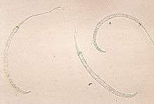
Dracunculus medinensis or Guinea worm is a nematode that causes dracunculiasis, also known as guinea worm disease.[1] The disease is caused by the female[2] which, at up to 80 cm (31 in) in length,[3] is among the longest nematodes infecting humans.[4] In contrast, the longest recorded male Guinea worm is only 4 cm (1.6 in).[3]
| Guinea worm | |
|---|---|
 | |
| Scientific classification | |
| Kingdom: | Animalia |
| Phylum: | |
| Class: | Chromadorea |
| Order: | Spirurida |
| Superfamily: | Dracunculoidea |
| Family: | Dracunculidae |
| Genus: | Dracunculus |
| Species: | D. medinensis |
| Binomial name | |
| Dracunculus medinensis (Linnaeus, 1758) | |
| Synonyms | |
|
Gordius medinensis Linnaeus, 1758 | |
The common name "guinea worm" is derived from the Guinea region of Western Africa.
History
Dracunculus medinensis (little dragon from Medina) was described in Egypt as early as the 15th century BC and possibly was the "fiery serpent" of the Israelites described in the Bible.[5]
In the mid-19th century, the nematode Camallanus lacustris, which infects freshwater fish, was discovered to develop in copepods. This led to the discovery in 1870 by Russian naturalist Alexei Fedchenko of the means of transmission of D. medinensis, via copepod intermediate hosts.[6]
Lifecycle
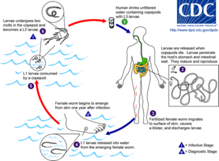
D. medinensis larvae are found in fresh water, where they are ingested by copepods of the genus Cyclops. Within the copepod, the D. medinensis larvae develop to an infective stage within 14 days.[7] When the infected copepod is ingested by a mammalian host, the copepod is dissolved by stomach acid and the D. medinensis larvae migrate through the wall of the mammalian intestine, and mature into adults. About 100 days after infection, a male and female D. medinensis meet and sexually reproduce within the host tissue. The male dies in the host tissue, while the female migrates to the host's subcutaneous tissue. Around a year after infection, the female causes the formation of a blister on the skin's surface, generally on the lower extremities, though occasionally on the hand or scrotum. When the blister ruptures, the female slowly emerges over the course of several days or weeks.[7] This causes extreme pain and irritation to the host. When the host submerges the affected body part in water, the female expels thousands of larvae into the water. From here, the larvae infect copepods, continuing the lifecycle.[7]
Animal reservoirs
As of 2016, more than 500 dogs in Chad, 13 in Ethiopia, and one dog in each of Mali and South Sudan, have been diagnosed with guinea worm.[8] Furthermore, it has been possible to infect frogs under laboratory conditions, and recently, natural infection has been reported in wild frogs in Chad.[9] These findings are a potential problem for the eradication program.
Epidemiology
D. medinensis is most commonly found in the subtropic to tropical regions, especially in India, south-west Asia (Iraq, Iran, Pakistan, etc.), and rural areas of Africa, where temperatures between 25 and 30 °C are best for larval development.[10] The parasite relies on people accidentally consuming microcrustaceans of the genus Cyclops (copepods), that dwell in stationary bodies of water such as ponds, large, open wells (with stairs), or rain-filled cisterns.[10] The infection occurs most during times of drought or the “dry-season” in humid climates, or during or just after the rain season in the “semiarid, wet-and-dry-climates.”[10] This is due to the lower surface water of the stationary bodies of water, which are prime for the growth of the infected copepods, and main source of water for many.[10]
Pathology
D. medinensis causes dracunculiasis as a result of the emergence of the female worm, nonemergence of adult worms (usually the male), and secondary bacterial infections.[11] As it emerges to the subcutaneous tissue, the female releases a toxic chemical that may result in nausea, rash at site, diarrhea, dizziness, localized edema, reddish papule, blister, and itching.[11] Arthritis or paraplegia can result from a worm that fails to reach the skin and gets calcified in or along the joint or finds its way into the central nervous tissue.[11] Aseptic abscesses and cystic swelling can also occur when worms rupture before emerging, causing an acute inflammatory response from the host's immune system.[11]
Treatment
The female guinea worm slowly starts to emerge from the host's skin after the blister ruptures. The most common method for removing the worm involves submerging the affected body part in water to help coax the worm out. The site is then cleaned thoroughly. Then, slight pressure is applied to the worm as it is slowly pulled out of the wound. To avoid breaking the worm, pulling should stop when resistance is met. Full extraction of the female guinea worm usually takes several days. After each day's worth of extraction, the exposed portion of the worm is wrapped around a piece of rolled-up gauze or small stick to maintain tension.[12] This method of wrapping the worm around a stick or gauze is speculated to be the source for the Rod of Asclepius, the symbol of medicine.[13] Once secure, topical antibiotics are applied to the affected region, to help prevent secondary infections due to bacteria, which is then wrapped in gauze to protect the wound. The same steps are repeated each day until the whole worm has been removed from the lesion.[12]
Eradication program
| Year | Reported cases | Countries |
|---|---|---|
| 1986 | 3,500,000 | 21 [15] |
| 1989 | 892,055 | 15 [16] |
| 1992 | 374,202 | 15 [16] |
| 1995 | 129,852 | 19 [16] |
| 2000 | 75,223 | 16 [16] |
| 2001 | 63,717 | 16 [16] |
| 2002 | 54,638 | 14 [16] |
| 2003 | 32,193 | 13 [16] |
| 2004 | 16,026 | 13 [16] |
| 2005 | 10,674 | 12 [16] |
| 2006 | 25,217 [lower-alpha 1] | 10 [16] |
| 2007 | 9,585 | 9 [16] |
| 2008 | 4,619 | 7 [16] |
| 2009 | 3,190 | 5 |
| 2010 | 1,797 | 4 [18] (6 [19]) |
| 2011 | 1,060 | 4 [20] |
| 2012 | 542 | 4 |
| 2013 | 148 | 5 |
| 2014 | 126 | 4 [21] |
| 2015 | 22 | 4 |
| 2016 | 25 | 3 [15] |
| 2017 | 30 | 2 [22] |
| 2018 | 28 | 3 [23] |
In the 1980s, the Carter Center initiated a program to eradicate guinea worm.[24] The campaign began in 1980 at the US Centers for Disease Control and Prevention. In 1984, the CDC was appointed as the World Health Organization Collaborating Center for research, training, and eradication of D. medinensis. More than twenty countries were affected by guinea worms in 1986. That year, WHO started the eradication program with the Carter Center leading the effort.[25] The program included education of people in affected areas that the disease was caused by larvae in drinking water, isolation and support for sufferers, and – crucially – widespread distribution of net filters and pipe filters for drinking water, and education about the importance of using them.
As of 2015, the species has been reported to be near eradication.[24] The International Commission for the Certification of Dracunculus Eradication has certified 198 countries, territories, and other WHO represented areas. In January 2015, eight countries remained to be certified as D. medinensis free. These eight countries include Angola, the Democratic Republic of the Congo, Kenya, Sudan, Chad, Ethiopia, Mali, and South Sudan. Of these, Chad, Ethiopia, Mali, and South Sudan are the only remaining endemic countries.[25] Not coincidentally, all four are affected by civil wars which affect the safety of health workers.
See also
- Planned extinction
Notes
- Increase in reported cases resulted from improved reporting in southern Sudan following the peace agreement in 2005.[17]
References
- Stefanie Knopp; Ignace K. Amegbo; David M. Hamm; Hartwig Schulz-Key; Meba Banla; Peter T. Soboslay (March 2008). "Antibody and cytokine responses in Dracunculus medinensis patients at distinct states of infection". Transactions of the Royal Society of Tropical Medicine and Hygiene. 102 (3): 277–283. doi:10.1016/j.trstmh.2007.12.003. PMID 18258273.
- Langbong Bimi (2007). "Potential vector species of Guinea worm (Dracunculus medinensis) in Northern Ghana". Vector-Borne and Zoonotic Diseases. 7 (3): 324–329. doi:10.1089/vbz.2006.0622. PMID 17767406.
- G. D. Schmidt; L S. Roberts (2009). Larry S. Roberts; John Janovy, Jr. (eds.). Foundations of Parasitology (8th ed.). McGraw-Hill. pp. 480–484. ISBN 978-0-07-128458-5.
- Talha Bin Saleem; Irfan Ahmed (2006). ""Serpent" in the breast" (PDF). Journal of Ayub Medical College Abbottabad. 18 (4): 67–68. PMID 17591014.
- "Dracunculiasis: Historical background". World Health Organization. Retrieved 27 April 2017.
- Roy C. Anderson (8 February 2000). Nematode Parasites of Vertebrates: Their Development and Transmission. CABI. p. 1. ISBN 978-0-85199-786-5.
- "Dracunculiasis: About Guinea-Worm Disease". World Health Organization. Retrieved 20 December 2015.
- "Dracunculiasis (guinea-worm disease)". World Health Organization. Retrieved 2016-10-02.
- Eberhard, Mark L.; Cleveland, Christopher A.; Zirimwabagabo, Hubert; Yabsley, Michael J.; Ouakou, Philippe Tchindebet; Ruiz-Tiben, Ernesto (2016). "Guinea Worm (Dracunculus medinensis) Infection in a Wild-Caught Frog, Chad". Emerging Infectious Diseases. 22 (11): 1961–1962. doi:10.3201/eid2211.161332. PMC 5088019. PMID 27560598.
- Muller, R. (1979). "Guinea worm disease: epidemiology, control, and treatment". Bulletin of the World Health Organization. 57 (5): 683–689. PMC 2395878. PMID 161522.
- Roberts, Larry S.; Janovy, John, Jr.; Nadler, Steve (2012-11-27). Gerald D. Schmidt & Larry S. Roberts' foundations of parasitology (9th ed.). New York. ISBN 9780073524191. OCLC 812614125.
- "Management of Guinea Worm Disease (GWD)". Parasites – Guinea Worm. Centers for Disease Control and Prevention. 2018-03-16. Retrieved 2018-07-22.
- Satin, Morton (2007). Death in the Pot: The Impact of Food Poisoning on History. Amherst: Prometheus Books. pp. 63–65.
- "Guinea Worm Case Totals". The Carter Center.
- Jeff Martin (January 11, 2017). "Carter: Guinea Worm disease reported in 3 countries in 2016". Associated Press. Retrieved 2017-01-11.
- "Dracunculiasis Epidemiological Data (1989-2008)" (PDF). World Health Organization. Retrieved 2009-09-14.
- "Progress Toward Global Eradication of Dracunculiasis, January 2005--May 2007". MMWR. 56 (32): 813–817. August 17, 2007. Retrieved 2011-03-16.
- "Guinea Worm Countdown: The Road to Eradication". The Carter Center. Archived from the original on 2011-09-06. Retrieved 2018-07-22.
- "Monthly report on dracunculiasis cases, January–December 2010" (PDF). Weekly Epidemiology Record. WHO. 86 (10): 92. 4 March 2011. Retrieved 2011-03-04.
- WHO Collaborating Center for Research, Training and Eradication of Dracunculiasis (January 9, 2012). "Guinea Worm Wrap-up #209" (PDF) – via The Carter Center.
- "Carter Center: 126 Cases of Guinea Worm Disease Remain Worldwide". The Carter Center. January 11, 2015. Retrieved 2015-05-09.
- "Announcement for 2017 Guinea Worm Disease Case Totals". The Carter Center (Press release). January 19, 2018. Archived from the original on 2018-01-20. Retrieved 2018-07-22.
- "28 Guinea Worm Cases Reported in 2018".
- World Science Festival (January 23, 2015). "This Species Is Close to Extinction and That's a Good Thing". TIME.
- "Eradication Program". Parasites – Guinea Worm. Centers for Disease Control and Prevention. 2017-05-02. Retrieved 2018-07-22.
External links

- "Donors Commit $240 Million to Fight Neglected Diseases". Archived from the original on 2014-04-04. Retrieved 2014-04-04.
- How to Slay a Dragon – documentary by Clifford Bestall, broadcast on Al Jazeera English, Spring 2014 (video, 47 min.)