Digital pathology
Digital pathology is a sub-field of pathology that focuses on data management based on information generated from digitized specimen slides. Through the use of computer-based technology, digital pathology utilizes virtual microscopy. Glass slides are converted into digital slides that can be viewed, managed, shared and analyzed on a computer monitor. With the practice of Whole-Slide Imaging (WSI), which is another name for virtual microscopy,[1] the field of digital pathology is growing and has applications in diagnostic medicine, with the goal of achieving efficient and cheaper diagnoses, prognosis, and prediction of diseases.
History
The roots of digital pathology go back to the 1960s, when first telepathology experiments took place. Later in the 1990s the principle of virtual microscopy[2] appeared in several life science research areas. At the turn of the century the scientific community more and more agreed on the term ”digital pathology” to denote digitization efforts in pathology. However in 2000 the technical requirements (scanner, storage, network) were still a limited factor for a broad dissemination of digital pathology concepts. Over the last 5 years this changed as new powerful and affordable scanner technology as well as mass / cloud storage technologies appeared on the market. The field of Radiology has undergone the digital transformation almost 15 years ago, not because radiology is more advanced, but there are fundamental differences between digital images in radiology and digital pathology: The image source in radiology is the (alive) patient, and today in most cases the image is even primarily captured in digital format. In pathology the scanning is done from preserved and processed specimens, for retrospective studies even from slides stored in a biobank. Besides this difference in pre-analytics and metadata content, the required storage in digital pathology is two to three orders of magnitude higher than in radiology. However, the advantages anticipated through digital pathology are similar to those in radiology:
- Capability to transmit digital slides over distances quickly, which enables telepathology scenarios.
- Capability to access past specimen from the same patients and/or similar cases for comparison and review, with much less effort then retrieving slides from the archive shelfs.
- Capability to compare different areas of multiple slides simultaneously (slide by slide mode) with the help of a virtual microscope.
- Capability to annotate areas directly in the slide and share this for teaching and research.
Digital pathology is today widely used for educational purposes[3] in telepathology and teleconsultation as well as in research projects. Digital pathology allows to share and annotate slides in a much easier way and to download annotated lecture sets generates new opportunities for e-learning and knowledge sharing in pathology. Digital pathology in diagnostics is an emerging and upcoming field.
Environment
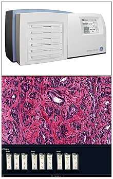
Scan
Digital slides are created from glass slides using specialized scanning machines. All high quality scans must be free of dust, scratches, and other obstructions.[4] These machines can be used to digitize slides and potentially train computers in deep learning (DL) exercises, to later preform pattern recognition-based tasks.
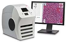
View
Digital slides are accessible for viewing via a computer monitor and viewing software either locally or remotely via the Internet.
Example: digital pathology tissue slide stained with Her2/neu biomarker used for diagnosis of breast cancer.
Manage
Digital slides are maintained in an information management system that allows for archival and intelligent retrieval.
Network
Digital slides are often stored and delivered over the Internet or private networks, for viewing and consultation.
Analyze
Image analysis tools are used to derive objective quantification measures from digital slides. Image segmentation and classification algorithms are used to identify medically significant regions and objects on digital slides. Recent developments in machine learning (ML) using deep learning methods have also emerged to make information hidden in integrated pathological data (images, patient history and -omics data) in arbitrarily high-dimensional spaces, both accessible and quantifiable. Thus, generating a novel source of information which is not yet available to the expert and not exploited in current Digital Pathology settings.
Integrate
Digital pathology workflow is integrated into the institution's overall operational environment. Slide digitization is expected to reduce the number of routine, manually reviewed slides, maximizing workload efficiency.
Sharing
Digital pathology also allows internet information sharing for education, diagnostics, publication and research. This may take the form of publicly available datasets or open source access to machine learning algorithms.
Challenges
Digital pathology has been approved by the FDA for primary diagnosis.[5] The approval was based on a multi-center study of 1,992 cases in which whole-slide imaging (WSI) was shown to be non-inferior to microscopy across a wide range of surgical pathology specimens, sample types and stains.[6] While there are advantages to WSI when creating digital data from glass slides, when it comes to real-time telepathology applications, WSI is not a strong choice for discussion and collaboration between multiple remote pathologists.[7] Furthermore, unlike digital radiology where the elimination of film made return on investment (ROI) clear, the ROI on digital pathology equipment is less obvious. The strongest ROI justification includes improved quality of healthcare, increased efficiency for pathologists, and reduced costs in handling glass slides.[8]
Potential
Trained pathologists traditionally view tissue slides under a microscope. These tissue slides may be stained to highlight cellular structures. When slides are digitized, they are able to be shared through tele-pathology and are numerically analyzed using computer algorithms. Algorithms can be used to automate the manual counting of structures, or for classifying the condition of tissue such as is used in grading tumors. They can additionally be used for feature detection of mitotic figures, epithelial cells, or tissue specific structures such as lung cancer nodules, glomeruli, or vessels.[9] This has the potential to reduce human error and improve accuracy of diagnoses. Digital slides can be easily shared, increasing the potential for data usage in education as well as in consultations between expert pathologists.
See also
References
- "Whole Slide Imaging | MBF Bioscience". www.mbfbioscience.com. Retrieved 2019-12-02.
- Ferreira, R; Moon, J; Humphries, J; Sussman, A; Saltz, J; Miller, R; Demarzo, A (1997). "The virtual microscope". Romanian Journal of Morphology and Embryology. 45: 449–453. PMC 2233368. PMID 9357666.
- Hamilton, Peter W.; Wang, Yinhai; McCullough, Stephen J.; Sussman (2012). "Virtual microscopy and digital pathology in training and education". APMIS. 120 (4): 305–315. doi:10.1111/j.1600-0463.2011.02869.x. PMID 22429213.
- Flagship Biosciences. "How to Improve Whole Slide Scanning in Digital Pathology". Flagship Biosciences LLC. Retrieved 25 September 2013.
- "FDA allows marketing of first whole slide imaging system for digital pathology". Retrieved May 24, 2017. Cite journal requires
|journal=(help) - Mukhopadhyay, Sanjay; Feldman, Michael; Abels, Esther (2017). "Whole slide imaging versus microscopy for primary diagnosis in surgical pathology: a multicenter randomized blinded noninferiority study of 1992 cases (pivotal study)". American Journal of Surgical Pathology. Epub ahead of print (1): 39–52. doi:10.1097/PAS.0000000000000948. PMC 5737464. PMID 28961557.
- Siegel, Gabriel; Regelman, Dan; Maronpot, Robert; Rosenstock, Moti; Hayashi, Shim-mo; Nyska, Abraham (Oct 2018). "Utilizing novel telepathology system in preclinical studies and peer review". Journal of Toxicologic Pathology. 31 (4): 315–319. doi:10.1293/tox.2018-0032. PMC 6206289. PMID 30393436.
- "How to Build a Business Case to Justify the Investment in Digital Pathology". Sectra Medical Systems. Retrieved April 26, 2015.
- Aeffner, Famke; Zarella, Mark D.; Buchbinder, Nathan; Bui, Marilyn M.; Goodman, Matthew R.; Hartman, Douglas J.; Lujan, Giovanni M.; Molani, Mariam A.; Parwani, Anil V.; Lillard, Kate; Turner, Oliver C. (2019-03-08). "Introduction to Digital Image Analysis in Whole-slide Imaging: A White Paper from the Digital Pathology Association". Journal of Pathology Informatics. 10. doi:10.4103/jpi.jpi_82_18. ISSN 2229-5089. PMC 6437786. PMID 30984469.
Further reading
- Kayser, K; Kayser, G; Radziszowski, D; Oehmann, A (1999). "From telepathology to virtual pathology institution: The new world of digital pathology" (PDF). Romanian Journal of Morphology and Embryology. 45: 3–9. PMID 15847374.
- McCullough, Bruce; Ying, Xiaoyou; Monticello, Thomas; Bonnefoi, Marc (2004). "Digital Microscopy Imaging and New Approaches in Toxicologic Pathology". Toxicologic Pathology. 32 (5): 49–58. doi:10.1080/01926230490451734. PMID 15503664.
- Schlangen, David; Stede, Manfred; Bontas, Elena Paslaru (2004). "Feeding OWL: Extracting and Representing the Content of Pathology Reports". NLPXML '04 Proceedings of the Workshop on NLP and XML. Nlpxml '04: 43–50.
- Cruz-Roa, Angel; Díaz, Gloria; Romero, Eduardo; González, Fabio (2011). "Automatic Annotation of Histopathological Images Using a Latent Topic Model Based On Non-negative Matrix Factorization". Journal of Pathology Informatics. 2 (4): 4. doi:10.4103/2153-3539.92031. PMC 3312710. PMID 22811960.
- "E-Health and Telemedicine". International Journal of Computer Assisted Radiology and Surgery. 1 (Supplement 1): 119–35. 2006. doi:10.1007/s11548-006-0012-1.
- Fine, Jeffrey L.; Grzybicki, Dana M.; Silowash, Russell; Ho, Jonhan; Gilbertson, John R.; Anthony, Leslie; Wilson, Robb; Parwani, Anil V.; et al. (2008). "Evaluation of whole slide image immunohistochemistry interpretation in challenging prostate needle biopsies". Human Pathology. 39 (4): 564–72. doi:10.1016/j.humpath.2007.08.007. PMID 18234276.
- Kayser, Klaus; Kayser, Gian; Radziszowski, Dominik; Oehmann, Alexander (2004). "New Developments in Digital Pathology: from Telepathology to Virtual Pathology Laboratory". In Duplaga, Mariusz; Zieliński, Krzysztof; Ingram, David (eds.). Transformation of Healthcare with Information Technologies. Studies in Health Technology and Informatics. 105. IOS Press. pp. 61–9. ISBN 978-1-58603-438-2. ISSN 0926-9630. PMID 15718595.
- Tolksdorf, Robert; Bontas, Elena Paslaru (2004). "Organizing Knowledge in a Semantic Web for Pathology". Object-Oriented and Internet-Based Technologies. Lecture Notes in Computer Science. 3263. pp. 115–56. doi:10.1007/978-3-540-30196-7_4. ISBN 978-3-540-23201-8.
- Potts, Steven J. (2009). "Digital pathology in drug discovery and development: Multisite integration". Drug Discovery Today. 14 (19–20): 935–41. doi:10.1016/j.drudis.2009.06.013. PMID 19596461.
- Potts, Steven J.; Young, G. David; Voelker, Frank A. (2010). "The role and impact of quantitative discovery pathology". Drug Discovery Today. 15 (21–22): 943–50. doi:10.1016/j.drudis.2010.09.001. PMID 20946967.
- Zwonitzer, R; Kalinski, T; Hofmann, H; Roessner, A; Bernarding, J (2007). "Digital pathology: DICOM-conform draft, testbed, and first results". Computer Methods and Programs in Biomedicine. 87 (3): 181–8. doi:10.1016/j.cmpb.2007.05.010. PMID 17618703.