Chorioallantoic membrane
The chorioallantoic membrane — also called the chorioallantois or abbreviated to CAM — is a vascular membrane found in eggs of some amniotes, such as birds and reptiles. It is formed by the fusion of the mesodermal layers of two developmental structures: the allantois and the chorion.[1] In mammals, this structure forms the placenta.
| Chorioallantoic membrane | |
|---|---|
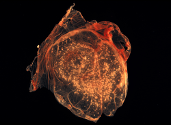 The chorioallantoic membrane of a developing chick covered with Smallpox virus pocks | |
| Identifiers | |
| MeSH | D049033 |
| Anatomical terminology | |
Three different layers compose the chorioallantoic membrane; these are called the chorionic epithelium, the mesenchyme and the allantoic epithelium. Blood capillaries and sinuses are found between epithelial cells of the chorionic layer, allowing close contact (within 0.2 μm) with air found in pores of the shell membrane of the egg.[2] As a result, the chorioallantoic membrane allows exchange of gases, such as oxygen, to developing embryos.[1] During embryonic development of birds, the chorioallantoic membrane also plays an essential role in bone formation by transporting calcium into the embryo from the eggshell.[1]
Chorioallantoic membranes from developing chicken eggs are routinely used in biological and biomedical research to investigate development,[3][4] angiogenesis,[5] tumors,[6][7] chemotherapeutic agents,[8] and to propagate and investigate viruses or helminths.[9][10][11]
References
- Gilbert, Scott F. (2003). Developmental biology. Sunderland, Mass: Sinauer Associates. ISBN 0-87893-258-5.
- Fáncsi T, Fehér G (June 1979). "Ultrastructural studies of chicken embryo chorioallantoic membrane during incubation". Anat Histol Embryol. 8 (2): 151–9. doi:10.1111/j.1439-0264.1979.tb00687.x. PMID 159001.
- Wilting J, Neeff H, Christ B (July 1999). "Embryonic lymphangiogenesis". Cell Tissue Res. 297 (1): 1–11. doi:10.1007/s004410051328. PMID 10398878.
- Osdoby P, Oursler MJ, Salino-Hugg T, Krukowski M (1988). "Osteoclast development: the cell surface and the bone environment". Ciba Found. Symp. 136: 108–24. PMID 3068005.
- Ribatti D (2008). "Chick embryo chorioallantoic membrane as a useful tool to study angiogenesis". Int Rev Cell Mol Biol. 270: 181–224. doi:10.1016/S1937-6448(08)01405-6. PMID 19081537.
- Cimpean AM, Ribatti D, Raica M (2008). "The chick embryo chorioallantoic membrane as a model to study tumor metastasis". Angiogenesis. 11 (4): 311–9. doi:10.1007/s10456-008-9117-1. PMID 18780151.
- Klingenberg, Marcel; Becker, Jürgen; Eberth, Sonja; Kube, Dieter; Wilting, Jörg (2014-05-18). "The chick chorioallantoic membrane as an in vivo xenograft model for Burkitt lymphoma". BMC Cancer. 14 (1). doi:10.1186/1471-2407-14-339. PMC 4036709. PMID 24884418.
- Klingenberg, Marcel; Becker, Jürgen; Eberth, Sonja; Kube, Dieter; Wilting, Jörg (2014-04-01). "The NADPH Oxidase Inhibitor Imipramine-Blue in the Treatment of Burkitt Lymphoma". Molecular Cancer Therapeutics. 13 (4): 833–841. doi:10.1158/1535-7163.MCT-13-0688. ISSN 1535-7163. PMID 24482381.
- Woolcock PR (2008). "Avian influenza virus isolation and propagation in chicken eggs". Methods Mol. Biol. 436: 35–46. doi:10.1007/978-1-59745-279-3_6. PMID 18370039.
- Guy JS (2008). "Isolation and propagation of coronaviruses in embryonated eggs". Methods Mol. Biol. 454: 109–17. doi:10.1007/978-1-59745-181-9_10. PMID 19057881.
- Fried B, Stableford LT (1991). "Cultivation of helminths in chick embryos". Adv. Parasitol. 30: 108–65. doi:10.1016/s0065-308x(08)60307-3. PMID 2069072.