Hydroxyapatite
Hydroxyapatite, also called hydroxylapatite (HA), is a naturally occurring mineral form of calcium apatite with the formula Ca5(PO4)3(OH), but it is usually written Ca10(PO4)6(OH)2 to denote that the crystal unit cell comprises two entities. Hydroxyapatite is the hydroxyl endmember of the complex apatite group. The OH− ion can be replaced by fluoride, chloride or carbonate, producing fluorapatite or chlorapatite. It crystallizes in the hexagonal crystal system. Pure hydroxyapatite powder is white. Naturally occurring apatites can, however, also have brown, yellow, or green colorations, comparable to the discolorations of dental fluorosis.
| Hydroxyapatite | |
|---|---|
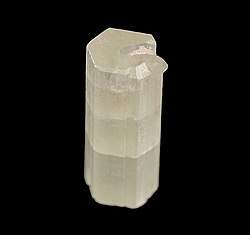
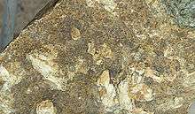 Hydroxylapatite crystals on matrix | |
| General | |
| Category | Phosphate mineral Apatite group |
| Formula (repeating unit) | Ca5(PO4)3(OH) |
| Strunz classification | 8.BN.05 |
| Crystal system | Hexagonal |
| Crystal class | Dipyramidal (6/m) H-M Symbol (6/m) |
| Space group | P63/m |
| Unit cell | a = 9.41 Å, c = 6.88 Å; Z = 2 |
| Identification | |
| Formula mass | 502.31 g/mol |
| Color | Colorless, white, gray, yellow, yellowish green |
| Crystal habit | As tabular crystals and as stalagmites, nodules, in crystalline to massive crusts |
| Cleavage | Poor on {0001} and {1010} |
| Fracture | Conchoidal |
| Tenacity | Brittle |
| Mohs scale hardness | 5 |
| Luster | Vitreous to subresinous, earthy |
| Streak | White |
| Diaphaneity | Transparent to translucent |
| Specific gravity | 3.14–3.21 (measured), 3.16 (calculated) |
| Optical properties | Uniaxial (-) |
| Refractive index | nω = 1.651 nε = 1.644 |
| Birefringence | δ = 0.007 |
| References | [1][2][3] |
Up to 50% by volume and 70% by weight of human bone is a modified form of hydroxyapatite, known as bone mineral.[4] Carbonated calcium-deficient hydroxyapatite is the main mineral of which dental enamel and dentin are composed. Hydroxyapatite crystals are also found in the small calcifications, within the pineal gland and other structures, known as corpora arenacea or 'brain sand'.[5]
Chemical synthesis
Hydroxyapatite can be synthesized via several methods, such as wet chemical deposition, biomimetic deposition, sol-gel route (wet-chemical precipitation) or electrodeposition.[6] Yagai and Aoki proposed the hydroxyapatite nanocrystal suspension can be prepared by a wet chemical precipitation reaction following the reaction equation below:[7]
10 Ca(OH)2 + 6 H3PO4 → Ca10(PO4)6(OH)2 + 18 H2O
Several studies have shown that hydroxyapatite synthesis via the wet-chemical route can be improved by high-power ultrasound. The ultrasonically assisted synthesis (sono-synthesis) of hydroxyapatite is a successful technique for the production of nanostructured hydroxyapatite to high quality standards. The ultrasonic route allows the production of nano-crystalline hydroxyapatite as well as modified particles, e.g. core-shell nanospheres and composites.[8]
Calcium deficient hydroxyapatite
Calcium deficient (non-stochiometric) hydroxyapatite, Ca10−x(PO4)6−x(HPO4)x(OH)2−x (where x is between 0 and 1) has a Ca/P ratio between 1.67 and 1.5. The Ca/P ratio is often used in the discussion of calcium phosphate phases.[9] Stoichiometric apatite Ca10(PO4)6(OH)2 has a Ca/P ratio of 10:6 normally expressed as 1.67. The non-stoichiometric phases have the hydroxyapatite structure with cation vacancies (Ca2+) and anion (OH–) vacancies. The sites occupied solely by phosphate anions in stochiometric hydroxyapatite, are occupied by phosphate or hydrogen phosphate, HPO42–, anions.[9] Preparation of these calcium deficient phases can be prepared by precipitation from a mixture of calcium nitrate and diammonium phosphate with the desired Ca/P ratio, for example to make a sample with a Ca/P ratio of 1.6:[10]
- 9.6 Ca(NO3)2 + 6 (NH4)2HPO4 → Ca9.6(PO4)5.6(HPO4)0.4(OH)1.6
Sintering these non-stoichiometric phases forms a solid phase which is an intimate mixture of tricalcium phosphate and hydroxyapatite, termed biphasic calcium phosphate:[11]
- Ca10−x(PO4)6−x(HPO4)x(OH)2−x → (1−x) Ca10(PO4)6(OH)2 + 3x Ca3(PO4)2
Biological function

The clubbing appendages of the Odontodactylus scyllarus (peacock mantis shrimp) are made of an extremely dense form of the mineral which has a higher specific strength and toughness than any synthetic composite material; these properties have led to its investigation for potential synthesis and engineering use.[12] Their dactyl appendages have excellent impact resistance due to the impact region being composed of mainly crystalline hydroxyapatite, which offers significant hardness. A periodic layer underneath the impact layer composed of hydroxyapatite with lower calcium and phosphorus content (thus resulting in a much lower modulus) inhibits crack growth by forcing new cracks to change directions. This periodic layer also reduces the energy transferred across both layers due to the large difference in modulus, even reflecting some of the incident energy.[13]
Hydroxyapatite is present in bone and teeth; bone is made primarily of HA crystals interspersed in a collagen matrix—65 to 70% of the mass of bone is HA. Similarly HA is 70 to 80% of the mass of dentin and enamel in teeth. In enamel, the matrix for HA is formed by amelogenins and enamelins instead of collagen.[14]
Hydroxylapatite deposits in tendons around joints results in the medical condition calcific tendinitis.[15]
Uses
Cosmetic
Hydroxyapatite is added to some variations of cornstarch based baby powder such as Johnson's Aloe & Vitamin E powder.[16] According the website, the mineral is added as an emollient to "help moisturize and soften skin."[17]
Medical
HA is increasingly used to make bone grafting materials as well as dental prosthetics and repair. Some implants, e.g. hip replacements, dental implants and bone conduction implants, are coated with HA.[14] As the native dissolution rate of hydroxyapatite in-vivo, around 10 wt% per year, is significantly lower than the growth rate of newly formed bone tissue, in its use as a bone replacement material, ways are being sought to enhance its solubility rate and thus promote better bioactivity.[18]
Supplement
Microcrystalline hydroxyapatite (MH) is marketed as a "bone-building" supplement with superior absorption in comparison to calcium.[19] It is a second-generation calcium supplement derived from bovine bone.[19] In the 1980s, bone meal calcium supplements were found to be contaminated with heavy metals,[19] and although the manufacturers claim their MH is free from contaminants, it isn't recommended because its effect in the body has not been well-tested.[19]
Chromatography
The mechanism of hydroxyapatite (HA) chromatography is complicated and has been described as "mixed-mode". It involves ionic interactions between positively charged groups on a biomolecule (often a protein) and the phosphate groups in hydroxyapatite, and metal chelation between hydroxyapatite calcium ions and negatively charged phosphate and/or carboxyl groups on the biomolecule. It may be difficult to predict the effectiveness of HA chromatography based on physical and chemical properties of the desired protein to be purified. For elution, a buffer with increasing phosphate and/or neutral salt concentration is typically used.
Use in archaeology
In archaeology, hydroxyapatite from human and animal remains can be analysed to reconstruct ancient diets, migrations and palaeoclimate. The mineral fractions of bone and teeth act as a reservoir of trace elements, including carbon, oxygen and strontium. Stable isotope analysis of human and faunal hydroxyapatite can be used to indicate whether a diet was predominantly terrestrial or marine in nature (carbon, strontium);[20] the geographical origin and migratory habits of an animal or human (oxygen, strontium)[21] and to reconstruct past temperatures and climate shifts (oxygen).[22] Post-depositional alteration of bone can contribute to the degradation of bone collagen, the protein required for stable isotope analysis.[23]
Defluoridation
Hydroxyapatite is a potential adsorbent for the defluoridation of drinking water as it forms fluorapatite in a 3 step process. In it, HAP updates F− from the water and replaces OH− ion to form the fluorapatite. However, during the defluoridation process the HAP dissolves, and increases the values of pH and phosphate ion, which makes the defluoridated water unfit for drinking. Recently, ″calcium amended-hydroxyapatite″ defluoridation technique is suggested to overcome the phosphate leaching from HAP. This technique also helps in the fluorosis reversal by providing the calcium-enriched alkaline drinking water to fluorosis affected areas.
See also
- Archaeological science
- Biomaterials: Mechanical Properties
- Calcium hydroxyphosphate
References
- Hydroxylapatite. Mindat
- Hydroxylapatite. Webmineral
- Anthony, John W.; Bideaux, Richard A.; Bladh, Kenneth W.; Nichols, Monte C., eds. (2000). "Hydroxylapatite". Handbook of Mineralogy (PDF). IV (Arsenates, Phosphates, Vanadates). Chantilly, VA, US: Mineralogical Society of America. ISBN 978-0962209734.
-
Junqueira, Luiz Carlos; José Carneiro (2003). Foltin, Janet; Lebowitz, Harriet; Boyle, Peter J. (eds.). Basic Histology, Text & Atlas (10th ed.). McGraw-Hill Companies. p. 144. ISBN 978-0-07-137829-1.
Inorganic matter represents about 50% of the dry weight of bone ... crystals show imperfections and are not identical to the hydroxyapatite found in the rock minerals
- Angervall, Lennart; Berger, Sven; Röckert, Hans (2009). "A Microradiographic and X-Ray Crystallographic Study of Calcium in the Pineal Body and in Intracranial Tumours". Acta Pathologica et Microbiologica Scandinavica. 44 (2): 113–119. doi:10.1111/j.1699-0463.1958.tb01060.x.
- Ferraz, M. P.; Monteiro, F. J.; Manuel, C. M. (2004). "Hydroxyapatite nanoparticles: A review of preparation methodologies". Journal of applied biomaterials & biomechanics : JABB. 2 (2): 74–80. PMID 20803440.
- Bouyer, E.; Gitzhofer, F.; Boulos, M. I. (2000). "Morphological study of hydroxyapatite nanocrystal suspension". Journal of Materials Science: Materials in Medicine. 11 (8): 523–31. doi:10.1023/A:1008918110156. PMID 15348004.
- Sono-Synthesis of Nano-Hydroxyapatite. hielscher.com
- Rey, C.; Combes, C.; Drouet, C.; Grossin, D. (2011). "1.111 – Bioactive Ceramics: Physical Chemistry". In Ducheyne, Paul (ed.). Comprehensive Biomaterials. 1. Elsevier. pp. 187–281. doi:10.1016/B978-0-08-055294-1.00178-1. ISBN 978-0-08-055294-1.
- Raynaud, S.; Champion, E.; Bernache-Assollant, D.; Thomas, P. (2002). "Calcium phosphate apatites with variable Ca/P atomic ratio I. Synthesis, characterisation and thermal stability of powders". Biomaterials. 23 (4): 1065–72. doi:10.1016/S0142-9612(01)00218-6. PMID 11791909.
- Valletregi, M. (1997). "Synthesis and characterisation of calcium deficient apatite". Solid State Ionics. 101–103: 1279–1285. doi:10.1016/S0167-2738(97)00213-0.
- Weaver, J. C.; Milliron, G. W.; Miserez, A.; Evans-Lutterodt, K.; Herrera, S.; Gallana, I.; Mershon, W. J.; Swanson, B.; Zavattieri, P.; Dimasi, E.; Kisailus, D. (2012). "The Stomatopod Dactyl Club: A Formidable Damage-Tolerant Biological Hammer". Science. 336 (6086): 1275–80. Bibcode:2012Sci...336.1275W. doi:10.1126/science.1218764. PMID 22679090.
- Tanner, K. E. (2012). "Small but Extremely Tough". Science. 336 (6086): 1237–8. Bibcode:2012Sci...336.1237T. doi:10.1126/science.1222642. PMID 22679085.
- Habibah, TU; Salisbury, HG (January 2018). "Biomaterials, Hydroxyapatite". PMID 30020686. Cite journal requires
|journal=(help) - Carcia, CR; Scibek, JS (March 2013). "Causation and management of calcific tendonitis and periarthritis". Current Opinion in Rheumatology. 25 (2): 204–9. doi:10.1097/bor.0b013e32835d4e85. PMID 23370373.
- https://www.johnsonsbaby.com/baby-products/johnsons-baby-powder-aloe-vitamin-e?upcean=381370052562
- https://www.johnsonsbaby.com/safety-standards/ingredients#calcium-hydroxyapatite
- Zhu, H.; et al. (2018). "Nanostructural insights into the dissolution behavior of Sr-doped hydroxyapatite". Journal of the European Ceramic Society. 38 (16): 5554–5562. doi:10.1016/j.jeurceramsoc.2018.07.056.
- Straub, D.A. (2007). "Calcium Supplementation in Clinical Practice: A Review of Forms, Doses, and Indications". Nutrition in Clinical Practice. 22 (3): 286–96. doi:10.1177/0115426507022003286. PMID 17507729.
- Richards, M. P.; Schulting, R. J.; Hedges, R. E. M. (2003). "Archaeology: Sharp shift in diet at onset of Neolithic" (PDF). Nature. 425 (6956): 366. Bibcode:2003Natur.425..366R. doi:10.1038/425366a. PMID 14508478.
- Britton, K.; Grimes, V.; Dau, J.; Richards, M. P. (2009). "Reconstructing faunal migrations using intra-tooth sampling and strontium and oxygen isotope analyses: A case study of modern caribou (Rangifer tarandus granti)". Journal of Archaeological Science. 36 (5): 1163–1172. doi:10.1016/j.jas.2009.01.003.
- Daniel Bryant, J.; Luz, B.; Froelich, P. N. (1994). "Oxygen isotopic composition of fossil horse tooth phosphate as a record of continental paleoclimate". Palaeogeography, Palaeoclimatology, Palaeoecology. 107 (3–4): 303–316. Bibcode:1994PPP...107..303D. doi:10.1016/0031-0182(94)90102-3.
- Van Klinken, G. J. (1999). "Bone Collagen Quality Indicators for Palaeodietary and Radiocarbon Measurements". Journal of Archaeological Science. 26 (6): 687–695. doi:10.1006/jasc.1998.0385.
External links
![]()
| Authority control |
|
|---|