Bulbus cordis
The bulbus cordis (the bulb of the heart) lies ventral to the primitive ventricle after the developing heart assumes its S-shaped form. Together, the bulbus cordis and the primitive ventricle give rise to the ventricles of the formed heart. The superior end of the bulbus cordis is also called the conotruncus.[1]
| Bulbus cordis | |
|---|---|
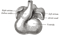 Heart showing expansion of the atria. | |
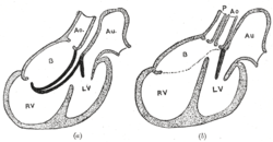 Diagrams to illustrate the transformation of the bulbus cordis. Ao. Truncus arteriosus. Au. Atrium. B. Bulbus cordis. RV. Right ventricle. LV. Left ventricle. P. Pulmonary artery. | |
| Details | |
| Carnegie stage | 9 |
| Gives rise to | smooth parts of right ventricle, left ventricle |
| Identifiers | |
| Latin | Bulbus cordis |
| Anatomical terminology | |
The adjacent walls of the bulbus cordis and ventricle approximate, fuse, and finally disappear, and the bulbus cordis now communicates freely with the right ventricle, while the junction of the bulbus with the truncus arteriosus is brought directly ventral to and applied to the atrial canal.
By the upgrowth of the ventricular septum the bulbus cordis is in great measure separated from the left ventricle, but remains an integral part of the right ventricle, of which it forms the infundibulum.
Additional images
 Head of chick embryo of about thirty-eight hours’ incubation, viewed from the ventral surface. X 26
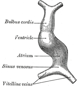 Diagram to illustrate the simple tubular condition of the heart.
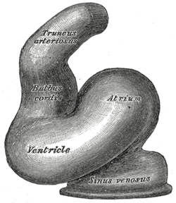 Heart of human embryo of about fourteen days.
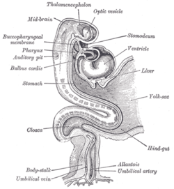 Human embryo about fifteen days old. Brain and heart represented from right side. Digestive tube and yolk sac in median section.
References
This article incorporates text in the public domain from page 513 of the 20th edition of Gray's Anatomy (1918)
- Larsen, William J (2001). Human Embryology (3rd ed.). Elsevier. p. 160. ISBN 0-443-06583-7.
External links
- Embryology at Temple Heart98/heart97b/sld023
- cardev-017—Embryo Images at University of North Carolina
- MedEd at Loyola GrossAnatomy/thorax0/Heart_Development/AtrioVent.html
- Kirby, Margaret L. (2007). Cardiac development. Oxford University Press. p. 119. ISBN 0-19-517819-X. Retrieved 20 April 2011.