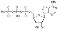
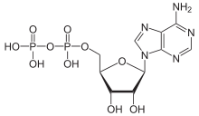
ATP hydrolysis
ATP hydrolysis is the catabolic reaction process by which chemical energy that has been stored in the high-energy phosphoanhydride bonds in adenosine triphosphate (ATP) is released by splitting these bonds, for example in muscles, by producing work in the form of mechanical energy. The product is adenosine diphosphate (ADP) and an inorganic phosphate, orthophosphate (Pi). ADP can be further hydrolyzed to give energy, adenosine monophosphate (AMP), and another orthophosphate (Pi).[1] ATP hydrolysis is the final link between the energy derived from food or sunlight and useful work such as muscle contraction, the establishment of electrochemical gradients across membranes, and biosynthetic processes necessary to maintain life.
The description and typical textbook labeling anhydridic bonds as "high energy . . bonds" can be very misleading to students. These bonds are in fact relatively weak. They do involve high energy electrons but the bonds themselves are quite easy to break. As noted below, energy is released by the hydrolysis of ATP when these weak bonds are broken – requiring a small input of energy, followed by the formation of new bonds and the release of a larger amount of energy as the total energy of the system is lowered and becomes more stable.[1]
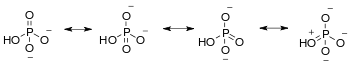
Hydrolysis of the phosphate groups in ATP is especially exergonic, because the resulting orthophosphate group is greatly stabilized by multiple resonance structures, making the products (ADP and Pi) much lower in energy than the reactant (ATP). The high negative charge density associated with the three adjacent phosphate units of ATP also destabilizes the molecule, making it higher in energy. Hydrolysis relieves some of these electrostatic repulsions, liberating useful energy in the process by causing conformational changes in enzyme structure.
In humans, approximately 60 percent of the energy released from the hydrolysis of ATP produces metabolic heat rather than fuel the actual reactions taking place.[2] Due to the acid-base properties of ATP, ADP, and inorganic phosphate, the hydrolysis of ATP has the effect of lowering the pH of the reaction medium. Under certain conditions, high levels of ATP hydrolysis can contribute to lactic acidosis.
How much energy ATP hydrolysis produces
Hydrolysis of the terminal phosphoanhydridic bond is a highly exergonic process. The amount of released energy depends on the conditions in a particular cell. Specifically, the energy released is dependent on concentrations of ATP, ADP and Pi. As the concentrations of these molecules deviate from values at equilibrium, the value of Gibbs free energy change (ΔG) will be increasingly different. In standard conditions ( ATP, ADP and Pi concentrations are equal to 1M, water concentration is equal to 55M) the value of ΔG is between -28 to -34 kJ/mol.[3][4]
The range of the ΔG value exists because this reaction is dependent on the concentration of Mg2+ cations, which stabilize the ATP molecule. The cellular environment also contributes to differences in the ΔG value since ATP hydrolysis is dependent not only on the studied cell, but also on the surrounding tissue and even the compartment within the cell. Variability in the ΔG values is therefore to be expected.[4]
The relationship between Gibbs free energy and chemical equilibrium is revealing. This relationship is defined by the equation ΔG=-RTln(Keq). The standard value of ΔG for this reaction is, as mentioned, between -28 and -34 kJ/mol; however, experimentally determined concentrations of the involved molecules reveal that the reaction is not at equilibrium.[4] Given this fact, a comparison between the equilibrium constant, K, and the reaction quotient, Q, provides insight. K takes into consideration reactions taking place in standard conditions, but in the cellular environment the concentrations of the involved molecules (namely, ATP, ADP, and Pi) are far from the standard 1M. In fact, the concentrations are more appropriately measured in mM, which is smaller than M by three orders of magnitude.[4] Using these nonstandard concentrations, the calculated value of Q is much less than one. By relating Q to ΔG using the equation ΔG=ΔG0+RTln(Q), where ΔG0 is the standard change in Gibbs free energy for the hydrolysis of ATP, the magnitude of ΔG is much greater than the standard. The nonstandard conditions of the cell actually result in a more favorable reaction.[5]
In one particular study, to determine the ΔG in vivo in humans, the concentration of ATP, ADP, and Pi was measured using nuclear magnetic resonance.[4] In human muscle cells at rest, the concentration of ATP was found to be around 4 mM and the concentration of ADP was around 9 μM. Inputing these values into the above equations yields the -64 kJ/mol ΔG. After ischemia, when the muscle is recovering from exercise, the concentration of ATP is as low as 1 mM and the concentration of ADP is around 7 μmol/l. Therefore, the absolute ΔG would be as high as -69 kJ/mol.[6]
By comparing the standard value of ΔG and the experimental value of ΔG, one can see that the energy released from the hydrolysis of ATP, as measured in humans, is almost twice as much as the energy produced in standard conditions.[4][5]
See also
- Dephosphorylation
References
- Syberg, F. Suveyzdis, Y., Kotting, C., Gerwert K., & Hofmann, E. Time-Resolved Fourier Transform Infrared Spectroscopy of the Nucleotide-binding Domain from the ATP-binding Cassette Transporter MsbA: ATP HYDROLYSIS ID THE RATE-LIMITING STEP IN THE CATALYTIC CYCLE. Journal of Biological Chemistry, 278(28), 23923-23931 doi:10.1074/jbc.M112.359208
- Zharova, T.V, & Vinogradov, Proton-Translocating ATP-synthase of Paracoccus denitrificans: ATP- Hydrolytic Activity. Biochemistry (00062979:, 68(10), A.D. (2003). 1101-1108
- Kamerlin, S. C., & Warshel, A. (2009). On the energetics of ATP hydrolysis in solution. The Journal of Physical Chemistry B, 113(47), 15692-15698.link to article
- Bergman, C., Kashiwaya, Y., & Veech, R. L. (2010) The effect of pH and Free Mg2+ on ATP Linked Enzymes and the Calculation of Gibbs Free Energy of ATP Hydrolysis. Journal of Physical Chemistry B, 114 (49), 16137-16146.
- Berg, J. M., Tymoczko, J. L., Stryer, L., Biochemistry, international edition. W. H. Freeman.; New York, New York, 2011; p 287
- Specific
- Lodish, Harvey (2013). Molecular cell biology (7th ed.). New York: W.H. Freeman and Co. pp. 52, 53. ISBN 9781464109812. OCLC 171110915.
- Berne & Levy physiology. Berne, Robert M., 1918-2001., Koeppen, Bruce M., Stanton, Bruce A. (6th, updated ed.). Philadelphia, PA: Mosby/Elsevier. 2010. ISBN 9780323073622. OCLC 435728438.CS1 maint: others (link)
- "Standard Gibbs free energy of ATP hydrolysis - Generic - BNID 101989". bionumbers.hms.harvard.edu. Retrieved 2018-01-25.
- Philips, Ron Milo & Ron. "» How much energy is released in ATP hydrolysis?". book.bionumbers.org. Retrieved 2018-01-25.
- "ATP: Adenosine Triphosphate". cnx.org. Retrieved 2018-05-16.
- Wackerhage, H.; Hoffmann, U.; Essfeld, D.; Leyk, D.; Mueller, K.; Zange, J. (December 1998). "Recovery of free ADP, Pi, and free energy of ATP hydrolysis in human skeletal muscle". Journal of Applied Physiology. 85 (6): 2140–2145. doi:10.1152/jappl.1998.85.6.2140. ISSN 8750-7587. PMID 9843537.