White matter dissection
White matter dissection refers to a special anatomical technique able to reveal the subcortical organization of white matter fibers in the human or animal cadaver brain.
The first studies of cerebral white matter (WM) were described by Galen and by the subsequent efforts of Vesalius on human cadaver specimens.[2][3] The interest for the deep anatomy of the brain pushed anatomist during centuries to create and develop different techniques for specimen preparation and dissection in order to better reveal the complex white matter architectural organization.[2][3][4][5]
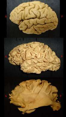
However, the biggest impact on the dissection of white matter anatomy was made by Joseph Klingler who developed a new method for specimens preparation and dissection. This technique became more feasible and widely used due to an increased quality of dissection and surprising quality of anatomical details.[4][5][6][7] Klingler developed a new method of brain fixation, by freezing already formalin-fixed brains before dissection.[6][7] First, the water crystallization induced by freezing disrupts the structure of the grey matter (which has a high water content). This process made possible to peel off the cortex from the brain surface without damaging the subcortical white matter organization underneath. Second, the freezing process along the WM fibers, induced a clear separation between them facilitating the dissection by progressive peeling of the fibers.[4][8][9]
White matter fibre dissection is nowadays considered as a valuable tool to enhance our knowledge about brain connectivity,[5][9][10][1] and has been used to validate tractographic results and vice versa with good consistency between the two techniques,[11] but also for neurosurgical training and neuroanatomical teaching.
References
- Latini F, Hjortberg M, Aldskogius H, Ryttlefors M, 2015. The Use of a Cerebral Perfusion and Immersion-Fixation Process for subsequent White Matter Dissection. J Neurosci Methods. Jul 3. pii: S0165-0270(15)00243-5. doi: 10.1016/j.jneumeth.2015.06.019
- Clarke E, O’Malley CD. 1968. The Human Brain and Spinal Cord, A Historical Study Illustrated By Writings from Antiquity To The Twentieth Century. Berkeley and Los Angeles: University of California Press.
- Clarke E, O’Malley CD. 1996. The Human Brain and Spinal Cord. A Historical Study Illustrated by Writings from Antiquity to the Twentieth Century. San Francisco, CA: Norman Publishing
- Ture U, Yasargil MG, Friedman AH, et al. 2000. Fiber dissection technique: lateral aspect of the brain. Neurosurgery 47: 417–427
- Agrawal A, Kapfhammer JP, Kress A, Wichers H, Deep A, Feindel W, Sontag VK, Spetzler RF, Preul MC. 2011. Josef Klingler's models of white matter tracts: influences on neuroanatomy, neurosurgery, and neuroimaging. Neurosurgery;69(2):238-52
- Klinger J. 1935. Erleichterung der makroskopischen Präparation des Gehirn durch den Gefrierprozess. Schweiz Arch-Neurol Psychiat 36: 247–256
- Ludwig E, Klinger J. 1956. Atlas Cerebri Humani. Basel: S. Karger.
- Fernandez-Miranda JC, Rhoton AL Jr, Alvarez-Linera J, Kakizawa Y, Choi C, de Oliveira EP. 2008. Three-dimensional microsurgical and tractographic anatomy of the white matter of the human brain. Neurosurgery 62:989-1026; discussion 1026– 1028.
- Zemmoura I, Blanchard E, Raynal PI, Rousselot-Denis C, Destrieux C, Velut S. 2015. How Klingler's dissection permits exploration of brain structural connectivity? An electron microscopy study of human white matter. Brain Struct Funct. Apr 24. DOI 10.1007/s00429-015-1050-7
- Arnts H, Kleinnijenhuis M, Kooloos JGM, Schepens-Franke AN, van Cappellen van Walsum AM. 2014. Combining fibre dissection, plastination and diffusion tensor imaging tractography for neuroanatomical education. Anatomical Sciences Education 7:47–55.
- Zemmoura I, Serres B, Andersson F, Barantin L, Tauber C, Filipiak I, Cottier JP, Venturini G, Destrieux C . 2014. FIBRASCAN: a novel method for 3D white matter tract reconstruction in MR space from cadaveric dissection. NeuroImage 103:106–118