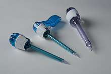
Trocar
A trocar (or trochar) is a medical or veterinary device that is made up of an obturator (which may be a metal or plastic sharpened or non-bladed tip), a cannula (essentially a hollow tube), and a seal. Trocars are placed through the abdomen during laparoscopic surgery. The trocar functions as a portal for the subsequent placement of other instruments, such as graspers, scissors, staplers, etc. Trocars also allow the escape of gas or fluid from organs within the body.
Etymology
The word trocar, less commonly trochar,[1] comes from French trocart, trois-quarts (three-fourths), from trois 'three' and carre 'side, face of an instrument',[2][3] first recorded in the Dictionnaire des Arts et des Sciences, 1694,[4] by Thomas Corneille, younger brother of Pierre Corneille.
History

Originally, doctors used trocars to relieve pressure build-up of fluids (edema) or gases (bloating). Patents for trocars appeared early in the 19th century, although their use dated back possibly thousands of years. By the middle of the 19th century, trocar-cannulas had become sophisticated, such as Reginald Southey's invention of the Southey tube.[5]
Applications
Medical/surgical use
Trocars are used in medicine to access and drain collections of fluid such as in a patient with hydrothorax or ascites.
In modern times, surgical trocars are used to perform laparoscopic (keyhole) surgery. They are deployed as a means of introduction for cameras and laparoscopic hand instruments, such as scissors, graspers, etc., to perform surgery hitherto carried out by making a large abdominal incision ("open" surgery), something that has revolutionized patient care. Today, surgical trocars are most commonly a single patient use instrument and have graduated from the "three-point" design that gave them their name to either a flat bladed "dilating-tip" product or something that is entirely blade free. This latter design offers greater patient safety due to the technique used to insert them.
Trocar insertion can lead to a perforating puncture wound of an underlying organ resulting in a medical complication. Thus, for instance, a laparoscopic intra-abdominal trocar insertion can lead to bowel injury leading to peritonitis or injury to large blood vessels with hemorrhage.[6]
Embalming
Trocars are also used near the end of the embalming process to provide drainage of bodily fluids and organs after the vascular replacement of blood with embalming chemicals. Rather than a round tube being inserted, the three sided knife of the classic trocar would split the outer skin into three "wings" which was then easily sutured closed in a less obtrusive way. It is attached to a suction hose (which usually is attached to a water aspirator). The process of removing gas, fluids, and semi-solids from the body cavities and hollow organs using the trocar is known as aspiration. The instrument is inserted into the body two inches to the left and two inches up from the navel. After the thoracic, abdominal, and pelvic cavities have been aspirated, the embalmer injects cavity fluid into the thoracic, abdominal and pelvic cavities using a smaller trocar attached via a hose which is connected to a bottle of high index cavity fluid. The bottle is held upside down in the air so as to let gravity take the cavity fluid through the trocar and into the cavities. The embalmer moves the trocar in the same manner used when aspirating the cavities in order to fully and evenly distribute the chemical.
After cavity embalming has been finished, the puncture is commonly sealed using a small, plastic object resembling a screw, called a trocar button.
Veterinary use
Trocars are widely used by veterinarians not only for draining hydrothorax, ascites, or for introducing instruments in laparoscopic surgery, but for acute animal-specific conditions as well. In cases of ruminal tympany (bloat) in cattle, a wide-bore trocar may be passed through the skin into the rumen to release trapped gas.[7] In dogs, a similar procedure is often performed for patients presenting with GDV (gastric dilatation-volvulus) in which a wide-bore trocar is passed through the skin into the stomach to immediately decompress the stomach. Depending on the severity of clinical signs on presentation, this is often performed after pain management has been administered but prior to general anaesthesia. Definitive surgical treatment involves anatomical repositioning of the stomach and spleen followed by a right-sided gastropexy.[8] Depending on the severity, partial gastrectomy and/or splenectomy may be indicated if the relevant tissues have necrosed due to ischemia caused by torsion/avulsion of the supplying vasculature. 72-hour post-operative supportive care, fluid therapy and ECG monitoring for cardiac arrhythmias is highly recommended.
Notes
- "trocar". Merriam-Webster Dictionary. Merriam-Webster. Retrieved December 6, 2018.
- "trocar". Oxford Dictionaries. Oxford University Press. Retrieved December 6, 2018.
- Kucklick, Theodore R. (2013). The Medical Device R&D Handbook (2 ed.). Boca Raton, FL: CRC Press, Taylor & Francis Group. p. 109. Retrieved December 6, 2018.
- Dictionnaire des Arts et des Sciences, de Thomas Corneille (1694, 1720, 1732)
- Library, Boston Medical (20 February 2018). "Boston Medical Library". www.countway.harvard.edu.
- S. Krishnakumar; P. Taube. "Entry Complications in Laparoscopic Surgery". J Gynecol Endosc Surg. 1: 4–11. doi:10.4103/0974-1216.51902. PMC 3304260. PMID 22442503.
- Constable, PD; Hinchcliff, KW; Done, SH; Gruenberg, W (2016). "Chapter 8: Diseases of the alimentary tract - ruminants. Ruminal tympany (bloat)". Veterinary Medicine: A textbook of the diseases of cattle, horses, sheep, pigs and goats (11 ed.). Elsevier Health Sciences. pp. 473–482. ISBN 9780702070587.
- Bright, Ronald M. (June 2007). "Acute Gastric Dilatation-Volvulus in Dogs" (PDF). Clinician's Brief.
References
- Janet Amundson Romich. An illustrated guide to veterinary medical terminology, Volume 1
- Mayer, Robert (2006). Embalming: History, Theory, and Practice (4th ed.). McGraw-Hill. ISBN 0-07-143950-1.