Triquetral bone
The triquetral bone (/traɪˈkwɛtrəl, -ˈkwiː-/; also called triquetrum, pyramidal, three-faced, and formerly cuneiform bone) is located in the wrist on the medial side of the proximal row of the carpus between the lunate and pisiform bones. It is on the ulnar side of the hand, but does not articulate with the ulna. It connects with the pisiform, hamate, and lunate bones. It is the 3rd most commonly fractured carpal bone.
| Triquetral bone | |
|---|---|
_01_palmar_view.png) Left hand anterior view (palmar view). Triquetral bone shown in red. | |
 The left triquetal bone | |
| Details | |
| Articulations | articulates with three bones: lunate laterally pisiform in front hamate distally triangular articular disk which separates it from the lower end of the ulna. |
| Identifiers | |
| Latin | os triquetrum, os pyramidale |
| MeSH | D051221 |
| TA | A02.4.08.006 |
| FMA | 23715 |
| Anatomical terms of bone | |
Structure
The triquetral is one of the eight carpal bones of the hand. It is a three-faced bone found within the proximal row of carpal bones. Situated beneath the pisiform, it is one of the carpal bones that form the carpal arch, within which lies the carpal tunnel. [1]:708
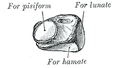
The triquetral bone may be distinguished by its pyramidal shape, and by an oval isolated facet for articulation with the pisiform bone. It is situated at the upper and ulnar side of the carpus. To facilitate its palpation in an exam, the hand must be radially deviated so that the triquetrium moves out from under the ulnar styloid process. The triquetrum may be difficult to find, since it also lies under the pisiform.
Ossification
The triquetral bone ossifies between 9 months and 50 months (4 years and 2 months).[2]
Surfaces
The inferior surface presents a medial, rough, non-articular portion, and a lateral convex articular portion which articulates with the triangular articular disk of the wrist.
The superior surface, directed lateralward, is concave, sinuously curved, and smooth for articulation with the hamate. The dorsal surface is rough for the attachment of ligaments.
The volar surface presents, on its medial part, an oval facet, for articulation with the pisiform; its lateral part is rough for ligamentous attachment.
The lateral surface, the base of the pyramid, is marked by a flat, quadrilateral facet, for articulation with the lunate.
The medial surface, the summit of the pyramid, is pointed and roughened, for the attachment of the ulnar collateral ligament of the wrist.
In animals
In reptiles and amphibians, the bone is instead referred to as the ulnare, since (at least in the most primitive fossils) it articulates with the ulna.
Fracture
Triquetral fractures can occur due to forceful flexion of the wrist, causing an avulsion of the dorsal aspect of the bone that is often hidden on anterior radiographs, but can be seen as a tiny bone fragment on lateral views.
Etymology
The etymology derives from the Latin triquetrus which means "three-cornered." Therefore, it is sometimes also called the triangular bone or os triangulare. However, os triangulare may also refer to a nearby accessory bone.
Additional images
_-_animation01.gif) Triquetral bone of the left hand (shown in red). Animation.
Triquetral bone of the left hand (shown in red). Animation._-_animation02.gif) Triquetral bone of the left hand. Close up. Animation.
Triquetral bone of the left hand. Close up. Animation. Triquetral bone.
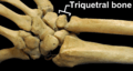 Right hand posterior view (dorsal view). Thumb on bottom.
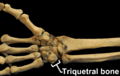 Right hand anterior view (palmar view). Thumb on top.
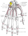 Bones of the left hand. Palmar surface. Triquetral shown in yellow.
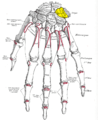 Bones of the left hand. Dorsal surface. Triquetral shown in yellow.
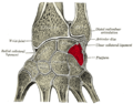 Cross section of wrist (thumb on left). Triquetral shown in red.
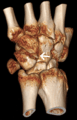 Triquetral fracture indicated by the white arrow.
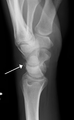 Triquetral fracture as seen on lateral view of a radiograph.
References
- Drake, Richard L.; Vogl, Wayne; Tibbitts, Adam W.M. Mitchell; illustrations by Richard; Richardson, Paul (2005). Gray's anatomy for students. Philadelphia: Elsevier/Churchill Livingstone. ISBN 978-0-8089-2306-0.
- Balachandran, Ajay; Kartha, Moumitha; Krishna, Anooj; Thomas, Jerry; K, Prathilash; TN, Prem; GK, Libu; B, Krishnan; John, Liza (2014). "A Study of Ossification of Capitate, Hamate, Triquetral & Lunate in Forensic Age Estimation". Indian Journal of Forensic Medicine & Toxicology. 8 (2): 218–224. doi:10.5958/0973-9130.2014.00720.8. ISSN 0973-9130. Retrieved 18 August 2014.